Showing posts with label Paediatrics. Show all posts

Tetralogy of Fallot


Tetralogy of Fallot

is the most common Cyanotic heart disease in children. The condition causes mixing of deoxygenated blood with the oxygenated blood being pumped out of the heart and into the circulatory system of blood vessels, Causing-

  • hypoxemia.

  • cyanosis, a bluish color of the skin, lips, and membranes inside the mouth and nose.
The 4 abnormalities (tetralogy) of the heart described by Fallot include the following:
  • Right ventricular hypertrophy: Narrowing or blockage of the pulmonary valve and/or muscle under the pulmonary valve coming out of the right ventricle. This restriction to blood outflow causes an increase in right ventricular work and pressure, leading to right ventricular thickening or hypertrophy.

  • Ventricular septal defect (VSD): This is a hole in the heart wall (septum) that separates the 2 ventricles. The hole is usually large and allows oxygen-poor blood in the right ventricle to pass through, mixing with oxygen-rich blood in the left ventricle. This poorly oxygenated blood is then pumped out of the left ventricle to the rest of the body. The body gets some oxygen, but not all that it needs. This lack of oxygen in the blood causes cyanosis.

  • Abnormal position of the aorta: The aorta, the main artery carrying blood out of the heart and into the circulatory system, exits the heart from a position overriding the right and left ventricles. (In the normal heart, the aorta exits from the left ventricle.) This is not of major importance in infants.

  • Pulmonary valve stenosis (PS): The major issue with tetralogy of Fallot is the degree of pulmonary valve stenosis, since VSD is always present. If the stenosis is mild, minimal cyanosis occurs, since blood still mostly travels to the lungs. However, if the PS is moderate to severe, a smaller amount of blood reaches the lungs, since most is shunted right-to-left through the VSD
Investigations:

CXR - baseline
ECG - yearly to assess rhythm and QRS duration
TTE - regularly (yearly or more frequent if severe PR and RV dysfunction, can be 2 or 3 yearly if very good surgical result and no new symptoms) (NB. It is easy to under estimate the degree of PR by colour follow alone) To assess pulmonary valve function, proximal PA anatomy, RV size and function, LV function, aortic root size and degree of aortic regurgitation.
Catheterisation - for dilatation / stenting of pulmonary arteries
MRI - becoming investigation of choice for proximal pulmonary artery anatomy, RV size and function and assessment of pulmonary regurgitation
Holter - for symptoms and screening for VT or high grade ectopy in those with marked RV dilatation or QRS > 180ms
Exercise test / CPEX - exercise capacity serial assessment, pre-pregnancy counselling

Additional investigations - EP studies for those with symptomatic atrial or ventricular arrhythmias or unexplained syncope or pre-syncope


Indications for intervention

Unrepaired tetralogy of Fallot (at any age)
Significant RVOT or branch PA stenosis (RV pressure > half systemic)
Severe pulmonary regurgitation with dilatation and dysfunction of the RV
Aortic regurgitation
Arrhythmias in presence of repairable haemodynamic sequela

TREATMENT OPTIONS
Surgery (+/- anti-arrhythmia procedures)

Percutaneous pulmonary valve implantation (for those with an RV to PA conduit of suitable size only)

Percutaneous pulmonary artery angioplasty and stenting

11 Categories Of Rickets

Because rickets results from a metabolic disturbance, the underlying disease should be diagnosed. The causes of rickets can be classified into 11 main categories:

  • Vitamin D deficiency
    • Dietary deficiency
    • Deficient endogenous synthesis
  • Gastrointestinal tract disorders
    • Small intestine diseases with malabsorption
    • Partial or total gastrectomy
    • Hepatobiliary disease
    • Chronic pancreatic insufficiency
  • Disorders of vitamin D metabolism
    • Hereditary - Pseudovitamin D deficiency or vitamin D dependency (types I and II)
    • Acquired
      • Use of anticonvulsants
      • Chronic renal failure
  • Acidosis
    • Distal renal tubular acidosis (classic or type I)
    • Secondary forms of renal acidosis
    • Ureterosigmoidostomy
    • Drug-induced disease
      • Chronic acetazolamine ingestion
      • Chronic ammonium chloride ingestion
  • Chronic renal failure
  • Phosphate depletion
    • Dietary - Low phosphate intake plus ingestion of nonabsorbable antacids
      • Hereditary - X-linked hypophosphatemic rickets or adult-onset vitamin D–resistant hypophosphatemic osteomalacia
      • Acquired - Sporadic hypophosphatemic osteomalacia (phosphate diabetes), tumor-associated (oncogenous) rickets, osteomalacia, neurofibromatosis, and fibrous dysplasia
  • Generalized renal tube disorders
    • Primary renal tube disorders
    • Renal tube disorders associated with systemic metabolic abnormality
      • Cystinosis
      • Glycogenosis
      • Lowe syndrome
    • Systemic disorder with associated renal disease
      • Hereditary - Inborn errors (Wilson disease, tyrosinemia) and neurofibromatosis
      • Acquired - Multiple myeloma, nephrotic syndrome, and kidney transplantation
      • Intoxication-related - Cadmium, lead, outdated tetracycline
  • Primary mineralization defects
    • Hereditary
    • Acquired
      • Diphosphonate treatment
      • Fluoride treatment
  • States of rapid bone formation with or without a relative defect in bone resorption
    • Postoperative hyperparathyroidism with osteitis fibrosa cystica
    • Osteopetrosis
  • Defective matrix synthesis - Fibrogenesis imperfecta ossium
  • Miscellaneous
    • Magnesium-dependent conditions
    • Axial osteomalacia
    • Parenteral alimentation
    • Aluminum intoxication
    • Isofosfamide treatment

Aishwarya Rai Syndrome and Hrithic Roshan Syndrome


Taken from New O.P. Ghai, 6th edition, page number 70,

It is a syndrome, given in O.P.Ghai,( possibility, may be ask the question in some entrances, at least in state PG)

" Issues regarding body weight,general attractiveness, breast size,complexion, and acne are some of the main body image concerns for adolescent girls."
It actually comes under Body Image Syndromes.

Similarly
Hrithic roshan Syndrome for Boys

Apert Syndrome : A genetic disorder

Apert Syndrome


Introduction

Apert syndrome is a rare genetic disorder that is characterized by specific craniofacial and limb abnormalities. It is caused by a genetic mutation in the FGFR2 gene on chromosome 10. The mutation can be inherited from a parent who has Apert syndrome or it can be a spontaneous (new) mutation. Studies show that Apert syndrome tends to occur more often in children with older fathers. Furthermore, all new mutations (those that have not been inherited by an affected parent) have been shown to occur exclusively in the FGFR2 gene received by the father. Apert syndrome occurs in 1 out of 100,000 to 160,000 live births and affects males and females equally. The first reported case of the syndrome was in 1848 by S.W. Wheaton, and in 1906, a French physician named E. Apert described nine cases and defined the syndrome.


Features and Characteristics

The following characteristics have been found in children with Apert syndrome:

  • Prematurely fused cranial sutures
  • Retruded (or sunken) mid-face
  • Fused fingers
  • Fused toes
  • Brachycephaly (short wide head)
  • Acrocephaly (high prominent forehead)
  • Flattened back of skull
  • Prominent eyes - may be spaced widely apart or slant downward
  • Strabismus
  • Prominent mandible
  • Depressed nasal bridge and small anteverted nose
  • Down-turned corners of the mouth
  • Low set ears (as well as hearing loss)
  • Cleft palate
  • Severe acne in teens
  • Hydrocephalus
  • Dental abnormalities (malposition of the teeth, crowding of the teeth, delayed tooth eruption, high-arched narrow palate, thickened ridges that support the teeth)
  • Internal organ abnormalities including heart defects and abnormalities of the trachea,
  • uterus, and brain
Skin manifestations of Apert syndrome
Hyperhidrosis
Synonychia
Brittle nails
Severe acne in puberty
Interruption of the eyebrows
Hypopigmentation
Hyperkeratosis
Paronychial infections of plantar skin
Excessive skin wrinkling of forehead
Dimples at knuckles, shoulders and elbows

Prematurely Fused Cranial Sutures

Retruded Mid-face

Fused Fingers and Toes

Diagnosis

The diagnosis can be made by a skull x-ray, which will confirm premature closure of the skull, and by a clinical exam. The combination of the craniofacial problems and the fused fingers and toes is what distinguishes Apert syndrome from other similar syndromes. Since the defect which causes Apert syndrome has been identified, genetic testing can be provided to confirm a diagnosis.

Treatment

Treating a child with Apert syndrome is best accomplished with a team approach. This would include a craniofacial surgeon, neurosurgeon, ENT specialist, audiologist, speech pathologist, oral surgeon, psychologist, ophthalmologist, and an orthodontist. The majority of treatment methods is surgical and the individual will likely require many operations. Aside from the surgeries required to correct the craniofacial problems and the fused fingers and toes, there may be other potential surgeries to improve the upper airway, address severe eye problems, or correct dental issues.
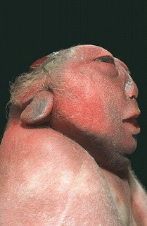
A child with Cushing syndrome

Anencephaly: Newborn Deformity

ANENCEPHALY
is a neural tube defect (a disorder involving incomplete development of the brain, spinal cord, and/or their protective coverings). The neural tube is a narrow sheath that folds and closes between the 3rd and 4th weeks of pregnancy to form the brain and spinal cord of the embryo. Anencephaly occurs when the "cephalic" or head end of the neural tube fails to close, resulting in the absence of a major portion of the brain, skull, and scalp. Infants with this disorder are born without both a forebrain (the front part of the brain) and a cerebrum (the thinking and coordinating area of the brain). The remaining brain tissue is often exposed--not covered by bone or skin. The infant is usually blind, deaf, unconscious, and unable to feel pain. Although some individuals with anencephaly may be born with a rudimentary brain stem, the lack of a functioning cerebrum permanently rules out the possibility of ever gaining consciousness. Reflex actions such as respiration (breathing) and responses to sound or touch may occur. The cause of anencephaly is unknown. Although it is believed that the mother's diet and vitamin intake may play a role, scientists believe that many other factors are also involved.

treatment

There is no cure or standard treatment for anencephaly. Treatment is supportive.

prognosis

The prognosis for individuals with anencephaly is extremely poor. If the infant is not stillborn, then he or she will usually die within a few hours or days after birth. [Editor's Note: The unborn child may have been diagnosed as having anencephaly, but be born with a less severe form of the disease, allowing the infant to live for years or more

SEARCH THIS BLOG FOR