Amyotrophic Lateral Sclerosis (ALS)

Amyotrophic Lateral Sclerosis (ALS)
Pop Star Jason beckeris surviving it.


Amyotrophic lateral sclerosis (ALS), also called Lou Gehrig's disease, is a disease that attacks the nerve cells (motor neurons) that control muscles. It gets worse over time (is progressive). Motor neurons carry messages about movement from the brain to the muscles, but in ALS the motor neurons degenerate and die; therefore, the messages to move no longer get to the muscles. When muscles aren't used for a long time, they weaken, waste away (atrophy), and twitch under the skin (fasciculate).
Eventually, all the muscles that a person can control (voluntary muscles) are affected. People with ALS lose the ability to move their arms, legs, mouth, and body. It may get to the point that the muscles used for breathing are affected, and the person might need a respirator (ventilator) in order to breathe. People with ALS can live 3 to 10 years or more after diagnosis.

Mental changesFor a long time it was believed that ALS only affected muscles. It is now known that one-third to one-half of individuals with ALS experience some changes in thinking (cognition). The disease can also cause changes in personality and behavior.


Who gets it?

People are most commonly diagnosed with ALS between the ages of 40 and 70, but younger people can also develop it. ALS affects people all over the world, in all ethnic backgrounds. Men are affected more often than women. About 90-95% of ALS cases appear at random, meaning no one in the person's family has the disorder. In about 5-10% of cases, a family member also has the disorder.


What causes it?

The exact cause of ALS is not known. In 1991, researchers identified a link between ALS and Chromosome 21. Two years later, a particular gene, SOD1, was identified as being associated with about 20% of the inherited cases in families. SOD1 controls an enzyme that breaks down free radicals, harmful particles that attacks cells from the inside and cause their death. Since not all inherited cases are connected to this gene, and some people are the only ones in their families with ALS, other genetic causes must exist.


Symptoms and diagnosis

Usually ALS comes on slowly, starting out as weakness in one or more muscles. Only one leg or arm may be affected. People notice that they stumble, having trouble lifting things, or have trouble with using their hands. As the disease progresses, the person with ALS will not be able to stand or walk, have trouble moving around, and trouble talking and swallowing. The diagnosis of ALS is based on the symptoms and signs the physician observes, as well as tests eliminating all the other possibilities, such as multiple sclerosis, post-polio syndrome, or infectious diseases.


Treatment

There is as yet no cure for ALS. Treatments are designed to relieve the symptoms and improve the quality of life for people with the disorder. Medications can help reduce fatigue, ease muscle cramps, and lessen pain. There is also a specific medication for ALS, called Rilutek (riluzole). It does not repair the damage already done to the body, but appears to be modestly effective in prolonging the survival of people with ALS. Through physical therapy, special equipment, and speech therapy, people with ALS can remain mobile and able to communicate.
LeaRN ABOUT bECKER AT www.xyzmusic.blogspot.com

Uterovaginal Prolapse: Types and Causes

Uterovaginal Prolapse Is a form of Hernia in which the uterus and Vagina protrudes down out of their normal confinement.

Types
I. Vaginal
a. Anterior wall
-Cystocele( Uriary bladder prolapse)
-Urethrocele( Urethra)
-Combined
b.Posterior wall
-Relaxed perinium
-Rectocele
-Vault prolapse

II. Uterine
Uterovaginal
Congenital

Causes Of Prolapse

Preciptating factors
I. Acquired
a. Overstretching of mackenrodth and Uterosacral Ligaments due to
Premature bear down
Application of forceps when cervix not fully dilated
down pressure on fundus to deliver placents
Precipitate labour
b. Overstretching of endopelvic fascial sheath of vagina
Degree of distension during delivery and duration
c.Subinvolution
ill nourished mother
early resumption of work
perisitent overfilling of bladder
repeated childbirth
d.Over stretching of perinium

Congenital
Spina Bifida occulta
Neurological disorders.

II. Aggravating factors.
a Increased abd pressure.
cough, constipation
b. bulky uterus, fibroid.
c.post menopausal atrophy
d.Asthenia and undernutrition


Overstretchin of perinium

Rhinophyma: Cosmetic Issue


Rhinophyma

Rhinophyma (an advanced type of rosacea) is characterized by an enlarged, bulbous, and red nose resulting from enlargement of the oil-producing glands beneath the surface of the skin on the nose. Depending on the severity of rhinophyma, doctors may begin treatment with topical or oral rosacea treatment.


What Is Rhinophyma?
To understand what rhinophyma is, it's important to first have a basic understanding of rosacea. Rosacea is a chronic disease that affects the skin. The disease is characterized by redness, pimples, and, in advanced stages, thickened skin. When rosacea reaches this advanced stage, rhinophyma may develop. The most common characteristic of rhinophyma is an enlarged, bulbous, and red nose. Rhinophyma is associated with the sebaceous (oil-producing) glands beneath the surface of the skin on the nose. The condition is more common in men than women.


Symptoms of Rhinophyma
Rhinophyma is characterized by an enlarged, bulbous, and red nose resulting from the enlargement of the sebaceous (oil-producing) glands beneath the surface of the skin on the nose.


What Causes Rhinophyma?
Doctors do not know the exact cause of rhinophyma.


Diagnosing Rhinophyma
Dermatologists usually diagnose rhinophyma by its appearance and associated symptoms because there are no tests that can diagnose this disease. However, on rare occasions, skin biopsies can pinpoint rhinophyma. Doctors will usually diagnose a patient with rhinophyma if the patient has rhinophyma symptoms and a family history of rhinophyma.


Current Treatment of Rhinophyma
Depending on the severity of the rhinophyma, doctors may begin treatment with topical or oral rosacea medications. Rhinophyma that does not respond to medications may be treated with:

Electrosurgery
Laser treatment
Dermabrasion
Resculpturing

ALOPECIA: HAIR LOSS causes


Hair loss


loss of hair is called alopecia.

Facts:
Hair loss usually develops gradually and may be patchy or diffuse (all over).

Roughly 100 hairs are lost from your head every day.

The average scalp contains about 100,000 hairs.
Each individual hair survives for an average of 4-1/2 years, during which time it grows about half an inch a month. Usually in its 5th year, the hair falls out and is replaced within 6 months by a new one.

Genetic baldness is caused by the body's failure to produce new hairs and not by excessive hair loss.
Both men and women tend to lose hair thickness and amount as they age. Inherited or "pattern baldness" affects many more men than women. About 25% of men begin to bald by the time they are 30 years old, and about two-thirds are either bald or have a balding pattern by age 60.


male pattern baldness involves a receding hairline and thinning around the crown with eventual bald spots. Ultimately, you may have only a horseshoe ring of hair around the sides. In addition to genes, male-pattern baldness seems to require the presence of the male hormone testosterone . Men who do not produce testosterone (because of genetic abnormalities or castration) do not develop this pattern of baldness.

Some women also develop a particular pattern of hair loss due to genetics, age, and male hormones that tend to increase in women after menopause. The pattern is different from that of men. Female pattern baldness involves a thinning throughout the scalp while the frontal hairline generally remains intact.

Common Causes:
Baldness is not usually caused by a disease, but is related to aging, heredity, and testosterone. In addition to the common male and female patterns from a combination of these factors, other possible causes of hair loss, especially if in an unusual pattern, include:
Alopecia areata -- bald patches that develop on the scalp, beard, and, possibly, eyebrows. Eyelashes may fall out as well.
Autoimmune conditions such as lupus
Burns
Certain infectious diseases such as syphilis
Chemotherapy
Emotional or physical stress
Excessive shampooing and blow-drying
Fever
Hormonal changes -- for example, thyroid disease , childbirth, or use of birth control pills
Nervous habits such as continual hair pulling or scalp rubbing
Radiation therapy
Tinea capitis (ringworm of the scalp)
Tumor of the ovary or adrenal glands

The Largest Physics experiment : Hoax of Doomsday ??


  1. When u attack the most difficult and fundamental questions [in science] you get a wide range of innovations, from MRI scanners to Facebook


"f you destroy particle physics and astronomy you will not produce more scientists working on carbon capture, you will just produce fewer scientists. "
Professor David L Wark


"The most powerful physics experiment ever built, the Large Hadron Collider will re-create the conditions just after the Big Bang in an attempt to answer fundamental questions of science and the universe itself. "


For More http://news.bbc.co.uk/2/hi/science/nature/7567926.stm


10 September,


News of Suicide by a Girl in India:


The Fear of Doomsday, Hoax made by Indian Cheap News Channels Like AAjtak, has lead to suicide of a Girl in Bhopal.


Since last 7-8 days the channels have been making a hoax of a doomsday resulting from this experiment, creating fear in the minds of the viewers. even my friends got scared... like these were the last few days to live...


Is it right to make such kinda publicites just to increase TPR of TV channels.?


Please leave you valuable coments>>>>


Birth defects Gallery

DOWN SYNDROME


HYDROCEPHALUS


ANENCEPHALY


Top Medical sites 2008

1 WebMD.com
6,742,178 - Inbound Links 11,264,170 - Compete Monthly Visitors 12,000,000 - Quantcast Monthly Visitors 1,653 - Alexa Ranking. Page Rank: 7Top 20 Health Websites Posted 08/19/2008 eBizMBA

2 NIH.gov
11,433,993 - Inbound Links 7,557,134 - Compete Monthly Visitors 9,000,000 - Quantcast Monthly Visitors 363 - Alexa Ranking. Page Rank: 9Top 20 Health Websites Posted 08/19/2008 eBizMBA

3 MayoClinic.com
409,085 - Inbound Links 3,751,224 - Compete Monthly Visitors 5,100,000 - Quantcast Monthly Visitors 3,792 - Alexa Ranking. Page Rank: 8Top 20 Health Websites Posted 08/19/2008 eBizMBA

4 MedicineNet.com
1,301,253 - Inbound Links 3,653,861 - Compete Monthly Visitors 4,600,000 - Quantcast Monthly Visitors 3,240 - Alexa Ranking. Page Rank: 7Top 20 Health Websites Posted 08/19/2008 eBizMBA

5 everydayHealth.com
12,535 - Inbound Links 4,771,021 - Compete Monthly Visitors 3,400,000 - Quantcast Monthly Visitors 12,764 - Alexa Ranking. Page Rank: 5Top 20 Health Websites Posted 08/19/2008 eBizMBA

6 Healthline.com
79,366 - Inbound Links 2,184,783 - Compete Monthly Visitors 3,000,000 - Quantcast Monthly Visitors 6,792 - Alexa Ranking. Page Rank: 6Top 20 Health Websites Posted 08/19/2008 eBizMBA

7 RevolutionHealth.com
765,016 - Inbound Links 2,103,307 - Compete Monthly Visitors 2,900,000 - Quantcast Monthly Visitors 13,344 - Alexa Ranking. Page Rank: 6Top 20 Health Websites Posted 08/19/2008 eBizMBA

8 Drugs.com
828,653 - Inbound Links 1,945,465 - Compete Monthly Visitors 2,300,000 - Quantcast Monthly Visitors 6,287 - Alexa Ranking. Page Rank: 6Top 20 Health Websites Posted 08/19/2008 eBizMBA

9 MedHelp.org
99,673 - Inbound Links 1,578,331 - Compete Monthly Visitors 2,000,000 - Quantcast Monthly Visitors 8,042 - Alexa Ranking. Page Rank: 6Top 20 Health Websites Posted 08/19/2008 eBizMBA

10 RxList.com
1,171,219 - Inbound Links 1,525,818 - Compete Monthly Visitors 2,000,000 - Quantcast Monthly Visitors 11,657 - Alexa Ranking. Page Rank: 7Top 20 Health Websites Posted 08/19/2008 eBizMBA

11 eMedicine.com
1,157,159 - Inbound Links 1,233,573 - Compete Monthly Visitors 1,500,000 - Quantcast Monthly Visitors 5,835 - Alexa Ranking. Page Rank: 7Top 20 Health Websites Posted 08/19/2008 eBizMBA

12 RealAge.com
41,051 - Inbound Links 1,577,068 - Compete Monthly Visitors 1,700,000 - Quantcast Monthly Visitors 8,852 - Alexa Ranking. Page Rank: 6Top 20 Health Websites Posted 08/19/2008 eBizMBA

13 QualityHealth.com
64,299 - Inbound Links 1,201,368 - Compete Monthly Visitors 1,500,000 - Quantcast Monthly Visitors 28,221 - Alexa Ranking. Page Rank: 5Top 20 Health Websites Posted 08/19/2008 eBizMBA

14 eDiets.com
136,820 - Inbound Links 1,238,852 - Compete Monthly Visitors 1,400,000 - Quantcast Monthly Visitors 8,239 - Alexa Ranking. Page Rank: 7Top 20 Health Websites Posted 08/19/2008 eBizMBA

15 familydoctor.org
474,004 - Inbound Links 856,565 - Compete Monthly Visitors 1,100,000 - Quantcast Monthly Visitors 12,655 - Alexa Ranking. Page Rank: NATop 20 Health Websites Posted 08/19/2008 eBizMBA

16 diet.com
119,873 - Inbound Links 1,160,260 - Compete Monthly Visitors 328,084 - Quantcast Monthly Visitors 10,629 - Alexa Ranking. Page Rank: 5Top 20 Health Websites Posted 08/19/2008 eBizMBA

17 Prevention.com
116,542 - Inbound Links 853,420 - Compete Monthly Visitors 685,947 - Quantcast Monthly Visitors 20,991 - Alexa Ranking. Page Rank: 6Top 20 Health Websites Posted 08/19/2008 eBizMBA

18 Healthology.com
2,774,943 - Inbound Links 580,139 - Compete Monthly Visitors 355,631 - Quantcast Monthly Visitors 32,541 - Alexa Ranking. Page Rank: 8Top 20 Health Websites Posted 08/19/2008 eBizMBA

19 MensHealth.com
180,139 - Inbound Links 439,530 - Compete Monthly Visitors 381,350 - Quantcast Monthly Visitors 7,744 - Alexa Ranking. Page Rank: 7Top 20 Health Websites Posted 08/19/2008 eBizMBA

20 WHO.int
1,848,622 - Inbound Links 205,802 - Compete Monthly Visitors NA - Quantcast Monthly Visitors 4,236 - Alexa Ranking. Page Rank: 8Top 20 Health Websites Posted 08/19/2008 eBizMBA

Chest Pain Differential Diagnosis

Myocardial Ischaemic Pain

The main feature of myocardial ischaemia (impending infarction) is usually prolonged chest pain. Typical characteristics of the pain include:
Duration usually over 20 minutes
Located in the retrosternal area, possibly radiating to the arms (usually to the left arm), back, neck, or the lower jaw
The pain is described as pressing or heavy or as a sensation of a tight band around the chest; breathing or changing posture does not notably influence the severity of the pain.
The pain is continuous, and its intensity does not alter
The symptoms (pain beginning in the upper abdomen, nausea) may resemble the symptoms of acute abdomen. Nausea and vomiting are sometimes the main symptoms, especially in inferoposterior wall ischaemia.
In inferoposterior wall ischaemia, vagal reflexes may cause bradycardia and hypotension, presenting as dizziness or fainting.
Electrocardiogram (ECG) is the key examination during the first 4 hours after pain onset, but normal ECG does not rule out an imminent infarction.
Markers of myocardial injury (cardiac troponins T and I, creatine kinase-MB mass) start to rise about 4 hours after pain onset. An increase of these markers is diagnostic of myocardial infarction irrespective of ECG findings.
Minor signs of myocardial infarction in ECG, see Table 1 in the original guideline document
Nonischaemic Causes of Chest Pain

Illness/condition Differentiating symptoms and signs
Reflux oesophagitis, oesophageal spasm
No ECG changes
Heartburn
Worse in recumbent position, but also while straining, like angina pectoris
The most common cause of chest pain

Pulmonary embolism
Tachypnoea, hypoxaemia, hypocarbia
No pulmonary congestion on chest x-ray
Clinical presentation may resemble hyperventilation.
Both arterial oxygen pressure (PaO2) and partial arterial pressure of carbon dioxide (PaCO2) decreased.
Pain is not often marked.
D-dimer assay positive

Hyperventilation
Hyperventilation Syndrome

The main symptom is dyspnoea, as in pulmonary embolism.
Often a young patient
Tingling and numbness of the limbs, dizziness
PaCO2 decreased, PaO2 increased or normal
Secondary Hyperventilation

Attributable to an organic illness/cause; acidosis, pulmonary embolism, pneumothorax, asthma, infarction, etc.

Spontaneous pneumothorax
Dyspnoea is the main symptom.
Auscultation and chest x-ray

Aortic dissection
Severe pain with changing localization
Type A dissection sometimes obstructs the origin of a coronary artery (usually the right) with signs of impending inferoposterior infarction
Pulses may be asymmetrical
Sometimes broad mediastinum on chest x-ray
New aortic valve regurgitation

Pericarditis
Change of posture and breathing influence the pain.
A friction sound may be heard.
ST-elevation but no reciprocal ST depression

Pleuritis
A stabbing pain when breathing. The most common cause of stabbing pain is, however, caused by prolonged cough

Costochondral pain
Palpation tenderness, movements of chest influence the pain
Might also be an insignificant incidental finding

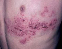
Early herpes zoster
No ECG changes, rash
Localized paraesthesia before rash

Ectopic beats
Transient, in the area of the apex

Peptic ulcer, cholecystitis, pancreatitis
Clinical examination (inferior wall ischaemia may resemble acute abdomen)

Depression
Continuous feeling of heaviness in the chest, no correlation to exercise
ECG normal

Alcohol-related
A young male patient in a casualty department, inebriated


ST changes resembling those of acute ischaemia
ST segment elevation
Early repolarization in V1–V3. Seen particularly in athletic men ("athlete's heart")
Acute myopericarditis in all leads except V1, aVR. Not resolved with a beta-blocker.
Pulmonary embolism – in inferior leads
Hyperkalaemia
Hypertrophic cardiomyopathy


ECG
ST segment depression
Sympathicotonia
Hyperventilation
Pulmonary embolism
Hypokalaemia
Digoxin
Antiarrhythmics
Psychiatric medication
Hypertrophic cardiomyopathy
Reciprocal ST depression of an inferior infarction in leads V2–V3–V4
Circulatory shock

QRS changes resembling those of Q wave infarction
Hypertrophic cardiomyopathy
Wolff-Parkinson-White (WPW) syndrome
Myocarditis
Blunt cardiac injury
Massive pulmonary embolism (QS in leads V1–V3)
Pneumothorax
Cardiac amyloidosis
Cardiac tumours
Progressing muscular dystrophy
Friedreich's ataxia

ST changes resembling those of a non-Q wave infarction
Increased intracranial pressure – subarachnoid bleed – skull injury
Hyperventilation syndrome
Post-tachyarrhythmia state
Circulatory shock – haemorrhage – sepsis
Acute pancreatitis
Myopericarditis

CAUSES OF ABDOMINAL PAIN: DD

Possible causes include:

Appendicitis (inflammation of the appendix)
Bowel obstruction
Cholecystitis (inflammation of the gallbladder) with or without gallstones
Chronic constipation
Dissecting abdominal aortic aneurysm
Diverticular disease, including diverticulitis
Easly-stage shingles (a viral infection where pain begins before the appearance of a rash)
Excessive gas
Food allergy
Food poisoning (salmonella, shigella)
Gastroesophageal reflux
Heartburn or indigestion
Hernia
Infectious mononucleosis
Inflammatory bowel disease (Crohn's disease or ulcerative colitis)
Intussusception -- while uncommon, this is a serious possible cause of pain in an infant who may be bringing the knees to the chest and crying
Irritable bowel syndrome
Kidney stones
Lactose intolerance
Pancreatitis (inflammation of the pancreas)
Parasitic infections (Giardia)
Sickle cell crisis
Spinal fracture
Ulcers
Urinary tract infections
Viral gastroenteritis (stomach flu)

Top causes Of cancer In USA.

FEMALES (total cases = 2,671,629)
Breast 30.4%
Lung 13.3%
Colon & Rectum 11.7%
Corpus & Uterus 5.7%
Non-Hodgkin Lymphoma 3.9%

MALES (total cases = 2,853,795)
Prostate 29.4%
Lung 15.8%
Colon & Rectum 11.2%
Bladder 6.5%
Non-Hodgkin Lymphoma 4.1%

Top 10 causes of Blindness

Causes of blindness and visual impairment

In spite of the progress made in surgical techniques in many countries during the last ten years, cataract (47.9%) remains the leading cause of visual impairment in all areas of the world, except for developed countries.
Other main causes of visual impairment in 2002 are
glaucoma (12.3%),
age-related macular degeneration (AMD) (8.7%),
corneal opacities (5.1%),
diabetic retinopathy (4.8%),
childhood blindness (3.9%),
trachoma (3.6%), and
onchocerciasis (0.8%).

The causes of avoidable visual impairment world wide are all the above except for AMD. In the least-developed countries, and in particular Sub-Saharan Africa, the causes of avoidable blindness are primarily, cataract (50%), glaucoma (15%), corneal opacities (10%), trachoma (6.8%), childhood blindness (5.3%) and onchocerciasis (4%).

Food Or Fuel? Option of the present World.

Biofuels brings food shortage and increase poverty.


A common objection to biomass energy production is that it could divert agricultural production away from food crops in a hungry world -- even leading to mass starvation in the poor countries.


The global rush to switch from oil to energy derived from plants will drive deforestation, push small farmers off the land and lead to serious food shortages and increased poverty unless carefully managed, says the most comprehensive survey yet completed of energy crops.The United Nations report, compiled by all 30 of the world organisation's agencies, points to crops like palm oil, maize, sugar cane, soya and jatropha. Rich countries want to see these extensively grown for fuel as a way to reduce their own climate changing emissions. Their production could help stabilise the price of oil, open up new markets and lead to higher commodity prices for the poor.
.....The UN warns: "Where crops are grown for energy purposes the use of large scale cropping could lead to significant biodiversity loss, soil erosion, and nutrient leaching. Even varied crops could have negative impacts if they replace wild forests or grasslands."
It' s Our ChoiceYou Need Food Or you Need Fuel???

Sudden Visual Loss or Blindness

Sudden Visual Loss

Etiology:
transient visual obscuration, TVO, papilledema, increased intracranial pressure, amaurosis fugax, monocular blindness, partial blindness, total blindness, transient monocular visual loss, TMVL, transient monocular blindness, TMB, transient bilateral visual loss, TBVL, ocular infarction, ischemic damage to the eye, internal carotid artery disease, ICA disease, angle-closure glaucoma, central retinal artery occlusion, CRAO, branch retinal artery occlusion, BRAO, ischemia of the optic nerve, anterior ischemic optic neuropathy, AION, ruptured globe, sudden visual loss, decreased vision, nonarteric anterior ischemic optic neuropathy, NAION, migraine, scintillating scotoma, intraocular foreign body, cardiac disease, stenotic vascular disease, carotid or vertebral artery atherosclerotic disease, fibromuscular dysplasia, arteritis, carotid artery dissection, vertebral artery dissection, platelet-containing emboli, hypercoagulable states, antiphospholipid syndrome, anemia

Prader Willi Syndrome: A short Review


Prader-Willi syndrome (Summarized By Me)


simplified


Prader-Willi syndrome (abbreviated PWS) is a very rare genetic disorder, in which seven genes (or some subset thereof) on chromosome 15 are missing or unexpressed (chromosome 15q partial deletion) on the paternal chromosome.


Diagnosis/testing
genetic testing, specifically DNA-based methylation testing to detect the absence of the paternally contributed Prader-Willi syndrome/Angelman syndrome (PWS/AS) region on chromosome 15q11-q13. Such testing detects over 97% of patients. Methylation-specific testing is important to confirm the diagnosis of PWS in all individuals, but especially those who are too young to manifest sufficient features to make the diagnosis on clinical grounds or in those individuals who have atypical findings.


PWS phenotype
Clinical Features


In Utero:
Reduced fetal movement
Frequent abnormal fetal position
At Birth:
Often breech or caesarean births
Lethargy
Hypotonia
Feeding difficulties (due to poor muscle tone affecting sucking reflex)
Difficulties establishing respiration
Hypogonadism
Infancy:
Failure to thrive (continued feeding difficulties)
Delayed milestones/intellectual delay
Excessive sleeping
Strabismus
Scoliosis (often not detected at birth)
Childhood:
Speech delay
Poor physical coordination
Hyperphagia (over-eating) from age 2 - 4 years. Note change from feeding difficulties in infancy
Excessive weight gain
Adolescence:
Delayed puberty
Short stature
Obesity
Extremely flexible
Adulthood:
Infertility (males and females)
Hypogonadism
Sparse pubic hair
Obesity
Hypotonia
Learning disabilities/borderline intellectual functioning (but some cases of average intelligence)
Proneness to diabetes mellitus
Extremely flexible
General physical appearance (adults)
Prominent nasal bridge
Small hands and feet
Soft skin, which is easily bruised
Excess fat, especially in the central portion of the body
High, narrow forehead
Almond shaped eyes with thin, down-turned lips
Light skin and hair relative to other family members
Lack of complete sexual development
Always picking at their skin
Stria
Delayed motor development


Neuro-cognitive
Individuals with PWS are at risk of learning and attention difficulties. Curfs and Frym (1992) conducted research into the varying degrees of learning disability found in Prader Willi Syndrome (PWS).[5] Their results were as follows:
5%...IQ above 85 (Average to low average intelligence)
27%..IQ 70 - 85 (Borderline intellectual functioning)
34%..IQ 50 - 70 (Mild intellectual disability)
27%..IQ 35 - 50 (Moderate intellectual disability)
5%...IQ 20 - 35 (Severe intellectual disability)
<1%..iq>


Behavioral
Prader-Willi syndrome is also frequently associated with an extreme and insatiable appetite, often resulting in morbid obesity. There is currently no consensus as to the cause for this particular symptom, although genetic abnormalities in chromosome 15 disrupt the normal functioning of the hypothalamus.[6] Given that the hypothalamus regulates many basic processes, including appetite, there may well be a link. However, no organic defect of the hypothalamus has been discovered on post mortem investigation.[6]


Endocrine
There are several aspects of PWS that support the concept of growth hormone deficiency in individuals with PWS. Specifically, individuals with PWS have short stature, are obese with abnormal body composition, have reduced fat free mass (FFM), have reduced LBM and total energy expenditure, and have decreased bone density.
PWS is characterized by hypogonadism. This is manifested as undescended testes in males and benign premature adrenarche in females. Testes may descend with time or can be managed with surgery or testosterone replacement. Adrenarche may be treated with hormone replacement therapy.


Treatment
Prader-Willi syndrome has no cure. However, several treatments are in place to lessen the condition's symptoms. Growth hormone replacement therapy improves body composition and increases linear height. During infancy, subjects should undergo therapies to improve muscle tone. Speech and occupational therapy are also indicated. During the school years, children benefit from a highly structured learning environment as well as extra help. Throughout their lives, the subject's food should literally be kept under lock and key, since the largest problem associated with the syndrome is severe obesity.
Because of severe obesity, obstructive sleep apnea is a common sequela, and a CPAP (continuous positive airway pressure) machine is often needed.


List Of All Infectious Diseases

Viral infectious diseases
AIDS – Chickenpox (Varicella) – Common cold – Cytomegalovirus Infection – Colorado tick fever – Dengue fever – Ebola hemorrhagic fever – Hand, foot and mouth disease – Hepatitis – Herpes simplex – Herpes zoster – HPV – Influenza (Flu) – Lassa fever – Measles – Marburg hemorrhagic fever – Infectious mononucleosis – Mumps – Norovirus – Poliomyelitis – Progressive multifocal leukencephalopathy – Rabies – Rubella – SARS – Smallpox (Variola) – Viral encephalitis – Viral gastroenteritis – Viral meningitis – Viral pneumonia – West Nile disease – Yellow feverBacterial Anthrax –

Bacterial
Meningitis –
Botulism – Brucellosis – Campylobacteriosis – Cat Scratch Disease – Cholera – Diphtheria – Epidemic Typhus – Gonorrhea – Impetigo– Legionellosis – Leprosy (Hansen's Disease) – Leptospirosis – Listeriosis – Lyme disease – Melioidosis – Rheumatic Fever;MRSA infection – Nocardiosis – Pertussis (Whooping Cough) – Plague – Pneumococcal pneumonia – Psittacosis – Q fever – Rocky Mountain Spotted Fever (RMSF) – Salmonellosis – Scarlet Fever – Shigellosis – Syphilis – Tetanus – Trachoma – Tuberculosis – Tularemia – Typhoid Fever – Typhus– Urinary Tract Infections

Parasitic infectious diseases
African trypanosomiasis – Amebiasis – Ascariasis – Babesiosis – Chagas Disease – Clonorchiasis – Cryptosporidiosis – Cysticercosis – Diphyllobothriasis – Dracunculiasis – Echinococcosis – Enterobiasis – Fascioliasis – Fasciolopsiasis – Filariasis – Free-living amebic infection – Giardiasis – Gnathostomiasis – Hymenolepiasis – Isosporiasis – Kala-azar – Leishmaniasis – Malaria – Metagonimiasis – Myiasis – Onchocerciasis – Pediculosis – Pinworm Infection – Scabies – Schistosomiasis – Taeniasis – Toxocariasis – Toxoplasmosis – Trichinellosis – Trichinosis – Trichuriasis – Trichomoniasis – Trypanosomiasis

Fungal infectious diseases
Aspergillosis – Blastomycosis – Candidiasis – Coccidioidomycosis – Cryptococcosis – Histoplasmosis – Tinea pedisPrion infectious diseases transmissible spongiform encephalopathy – Bovine spongiform encephalopathy – Creutzfeldt-Jakob disease – Kuru–Fatal Familial Insomnia–Alpers Syndrome –

Marfan Syndome: Tall is not always Gold

Marfan syndrome

is an autosomal dominant genetic disorder of the connective tissue characterized by disproportionately long limbs, long thin fingers, a typically tall stature, and a predisposition to cardiovascular abnormalities, specifically those affecting the heart valves and aorta. The disorder may also affect numerous other structures and organs — including the lungs, eyes, dural sac surrounding the spinal cord, and hard palate. It is named after Antoine Marfan, the French pediatrician

Symptoms

Three systems are predominantly affected.

Ocular:
upward lens dislocation
retinal detachment

Skeletal:
arachnodactyly
tall with disproportionately long legs and arms - the span of the arms is greater than the height
pectus excavatum
spinal abnormalities - spondylolisthesis, scoliosis
increased incidence of slipped upper femoral epiphysis
generalised joint laxity with predisposition to flat feet or dislocation of patella or shoulder

Cardiovascular
- affecting the aortic and mitral valves and the ascending aorta:
dilatation of the aorta may be noted at any age, beginning at the aortic valve and usually confined to the ascending aorta
aortic insufficiency may result from stretching of the aortic valve ring and a dissecting aneurysm of the aorta may be a terminal event. Rarely, it occurs during pregnancy
mitral insufficiency results from redundant cusps and chordae tendineae
other cardiac malformations have occasionally been reported
Mental development is normal.

The average lifespan of an affected individual is 40 to 50 years.


Skeletal system
The most readily visible signs are associated with the skeletal system. Many individuals with Marfan Syndrome grow to above average height. Some have long slender limbs with fingers and toes that are also abnormally long and slender (arachnodactyly). This long, slender body habitus and long, slender limbs are known as dolichostenomelia. An individual's arms may be disproportionately long, with thin, weak wrists. In addition to affecting height and limb proportions, Marfan syndrome can produce other skeletal signs. Abnormal curvature of the spine (scoliosis) is common, as is abnormal indentation (pectus excavatum) or protrusion (pectus carinatum) of the sternum. Other signs include abnormal joint flexibility, a high palate, malocclusions, flat feet, stooped shoulders, unexplained stretch marks on the skin and thin wrists. Some people with Marfan have speech disorders resulting from symptomatic high palates and small jaws.


Eyes
Marfan syndrome can also seriously affect the eyes and vision. Nearsightedness and astigmatism are common, but farsightedness can also result. Periodic eye exams can lead to an ophthalmologist or optometrist discovering dislocation, or subluxation, of the crystalline lens in one or both eyes (ectopia lentis) by carefully observing these structures using a slit-lamp biomicroscope. This can be differentiated from the similar condition homocystinuria, where the dislocation is inferonasal; in Marfan's the dislocation is superotemporal. Sometimes eye problems appear only after the weakening of connective tissue has caused detachment of the retina.Early onset glaucoma can be another complication.


Cardiovascular system
The most serious conditions associated with Marfan syndrome involve the cardiovascular system. Undue fatigue, shortness of breath, heart palpitations, racing heartbeats, or pain in the left chest, back, shoulder, or arm, can bring a person into the doctor's office. Cold arms, hands and feet can also be seriously linked to marfan syndrome because of a loss of blood circulation. A heart murmur heard on a stethoscope, an abnormal reading on an electrocardiogram, or symptoms of angina can lead a doctor to order an echocardiogram. This can reveal signs of leakage or prolapse of the mitral or aortic valves that control the flow of blood through the heart. (See mitral valve prolapse.) However, the major sign that would lead a doctor to consider an underlying condition is a dilated aorta or an aortic aneurysm. Sometimes, no heart problems are apparent until the weakening of the connective tissue in the ascending aorta causes an aortic aneurysm or even aortic dissection.


Lungs
Marfan syndrome is a risk factor for spontaneous pneumothorax. In spontaneous unilateral pneumothorax, air escapes from a lung and occupies the pleural space between the chest wall and a lung. The lung becomes partially compressed or collapsed. This can cause pain, shortness of breath, cyanosis, and, if not treated, death. Marfan syndrome has also been associated with sleep apnea and idiopathic obstructive lung disease.


Central nervous system
Another condition that can reduce the quality of life for an individual, though not life-threatening, is dural ectasia, the weakening of the connective tissue of the dural sac, the membrane that encases the spinal cord. Dural ectasia can be present for a long time without producing any noticeable symptoms. Symptoms that can occur are lower back pain, leg pain, abdominal pain, other neurological symptoms in the lower extremities, or headaches. Such symptoms usually diminish when the individual lies flat on his or her back. These types of symptoms might lead a doctor to order an X-ray of the lower spine. Dural ectasia is usually not visible on an X-ray in the early phases. A worsening of symptoms and the lack of finding any other cause should eventually lead a doctor to order an upright MRI of the lower spine. Dural ectasia that has progressed to the point of causing these symptoms would appear in an upright MRI image as a dilated pouch that is wearing away at the lumbar vertebrae. Other spinal issues associated with Marfan include degenerative disk disease and spinal cysts.

Sinusitis : A common cause of Headache

Sinusitis is an inflammation of the paranasal sinuses, which may or may not be as a result of infection, from bacterial, fungal, viral, allergic or autoimmune issues. Newer classifications of sinusitis refer to it as rhinosinusitis, taking into account the thought that inflammation of the sinuses cannot occur without some inflammation of the nose as well (rhinitis).

Types Of Sinusitis: ( BY LOCATION)

Maxillary sinusitis - can cause pain or pressure in the maxillary (cheek) area (e.g., toothache, headache) Frontal sinusitis - can cause pain or pressure in the frontal sinus cavity (located behind/above eyes), headache

Ethmoid sinusitis - can cause pain or pressure pain between/behind eyes, headache

Sphenoid sinusitis - can cause pain or pressure behind the eyes, but often refers to the vertex of the head

Acute sinusitis

Acute sinusitis is usually precipitated by an earlier upper respiratory tract infection, generally of viral origin. Virally damaged surface tissues are then colonized by bacteria, most commonly Haemophilus influenzae, Streptococcus pneumoniae, Moraxella catarrhalis and Staphylococcus aureus.
Other bacterial pathogens include other streptococci species, anaerobic bacteria and, less commonly, gram negative bacteria. Another possible cause of sinusitis can be dental problems that affect the maxillary sinus. Acute episodes of sinusitis can also result from fungal invasion. These infections are most often seen in patients with diabetes or other immune deficiencies (such as AIDS or transplant patients on anti-rejection medications) and can be life threatening. In type I diabetes, ketoacidosis causes sinusitis by Mucormycosis.
Chronic sinusitisChronic sinusitis is a complicated spectrum of diseases that share chronic inflammation of the sinuses in common. The causes are multifactorial and may include allergy, environmental factors such as dust or pollution, bacterial infection, or fungus (either allergic, infective, or reactive). Non allergic factors such as Vasomotor rhinitis can also cause chronic sinus problems. Abnormally narrow sinus passages, which can impede drainage from the sinus cavities, can also be a factor.
Symptoms include: Nasal congestion; facial pain; headache; fever; general malaise; thick green or yellow discharge; blurred vision, feeling of facial 'fullness' worsening on bending over; aching teeth.Very rarely, chronic sinusitis can lead to Anosmia, the inability to smell or detect odors.
A more recent, and still debated, development in chronic sinusitis is the role that fungus may play.
Predisposing Factors: deviated septum or small sinus ostia; smoking; nasal polyps; carrying the cystic fibrosis gene prior bouts of sinusitis

Treatment
Acute sinusitis
nasal irrigation or jala neti using a warm saline solution, hot drinks including tea and chicken soup, over-the-counter decongestants and nasal sprays, and getting plenty of rest. Analgesics (such as aspirin, paracetamol (acetaminophen) or ibuprofen) can be used, but caution must be employed to make sure the patient does not suffer from aspirin-exacerbated respiratory disease (AERD) as this could lead to anaphylaxis.
(Amoxicillin usually being the most common) with amoxicillin/clavulanate (Augmentin/Co-Amoxiclav) being indicated for patients who fail amoxicillin alone. Fluoroquinolones may be used in patients who are allergic to penicillinsDoxycycline is also used by some, for patients who are penicillin allergic

Chronic sinusitis
Simple measuresNasal irrigation and flush promotes sinus cavity z health, andpatients with chronic sinusitis including symptoms of facial pain, headache, halitosis, cough, anterior rhinorrhea (watery discharge) and nasal congestion found nasal irrigation to be "just as effective at treating these symptoms as the drug therapies." In other studies, "daily hypertonic saline nasal irrigation improves sinus-related quality of life, decreases symptoms, and decreases medication use in patients with frequent sinusitis," and is "recommended as an effective adjunctive treatment of chronic sinonasal symptoms.
Medical approaches
FESS - functional endoscopic sinus surgery, whereby normal clearance from the sinuses is restored by removing the anatomical and pathological obstructive variations that predispose to sinusitis. This replaces prior open techniques requiring facial or oral incisions and refocuses the technique to the natural openings of the sinuses instead of promoting drainage by gravity, the idea upon which the Caldwell-Luc surgery was based
balloon sinuplasty
Endoscopic nasal Surgery allows more functional approach than by radical antrostomy. Intranasal endoscopic operations permits minimal trauma to adjacent tissues and precise removal of the diseased mucosa. intranasal inferior meatal antrostomy middle meatal antrostomy Caldwell-Luc radical antrostomy
ComplicationsIntracranial Complications -"meningitis""abscess",
Orbital Complications Orbital CellulitisOrbital Abscess
Vascular Complications -Infections aneurysms or infected blood clots in the intracranial cavity, both of which are potentially fatal.
Asthma Loss of Smell and Taste - ngs. Osteomyelitis

Preventable Blindness

Causes:

Cataract

Corneal diseases

retinal pathology

glaucoma

trachoma

Injuries

Tonsillitis and Its Complications


Tonsillitis


is a disorder involving inflammation of the tonsils. There are two tonsils, situated on either side of the back of the throat and they form part of the body's immune system. They, like the rest of the immune system, contain special cells to trap and kill bacteria and viruses travelling through the body. When the main site of infection is within the tonsils they swell, become red and inflamed and may show a surface coating of white spots. Tonsillitis is extremely common in children and young people but it can occur at any age. The characteristics of the disease are pain in the throat and trouble swallowing.Tonsillitis is usually a self-limiting condition, ie it gets better without treatment, and generally there are no complications.


Cause

Whether due to viruses or bacteria, the infection is spread from person to person by airborne droplets, hand contact or kissing.There are many different individual viruses and bacteria that can potentially cause tonsillitis. For example the Epstein-Barr virus, which is the cause of glandular fever (infectious mononucleosis), is a common cause. It is typically seen in young people, particularly in situations such as university halls of residence when person-to-person spread is made easier by numerous close contacts. Among the bacteria that cause sore throats, the streptococcus group A is the most common - often known as 'strep throat'. The incubation period between picking up the infection and the disease breaking out is two to four days - sometimes it can be less.


signs and symptoms

Pain in the throat (sometimes severe) that may last more than 48 hours and be associated with difficulty in swallowing. The pain may spread to the ears.
The throat is reddened, the tonsils are swollen and may be coated or have white spots on them.
Possibly a high temperature.
Swollen lymph glands under the jaw and in the neck.
Headache.
Loss of voice or changes in the voice. If the sore throat is due to a viral infection the symptoms are usually milder and often related to the common cold. If due to Coxsackie virus infection, small blisters develop on the tonsils and roof of the mouth. The blisters erupt in a few days and are followed by a scab, which may be very painful.If the sore throat is due to a streptococcal infection, the tonsils often swell and become coated and the throat is sore. The patient has a temperature, foul-smelling breath and may feel quite ill.These different appearances are however very variable, and it is impossible to tell by looking at someone's throat whether the infection is due to a virus (which cannot be helped by antibiotics) or a bacterial infection (which might be helped by antibiotics). Good advice
If signs of a sore throat persist for more than a few days or are severe with marked difficulty in swallowing, high fever or vomiting, then you should consult your GP.
Warm drinks, soft food and the use of throat lozenges and/or a mouthwash may ease swallowing problems. Your pharmacist can advise on the most appropriate over-the-counter remedies.
Drink plenty of fluids. Understandably a sore throat can discourage swallowing but in tonsillitis it is common to lose a lot of body fluid through fever and mouth breathing. Dehydration adds considerably to feeling unwell.
It is important to ensure you have adequate rest and stay in a warm environment. How does the doctor make a diagnosis?The doctor usually makes the diagnosis from the symptoms and signs of the disease, but occasionally a swab of the secretions of the throat and maybe a blood sample are required to identify the cause. What complications may arise?Usually a throat infection such as tonsillitis causes no trouble and only lasts about a week, but the following complications can arise.


COPMPLICATIONS

A secondary infection may occur in the middle ear or sinuses.
If the sore throat is due to a streptococcus infection, there may be a rash (scarlet fever).
An uncommon complication is a throat abscess which occurs usually only on one side. If sufficiently large this can need surgical drainage (Quinsy throat).
In very rare cases, diseases like rheumatic fever or a particular kidney disease (glomerulonephritis) can occur. This is much less commonly observed now than it was several decades ago.


Treatment:

the vast majority of people, infection caused by a virus infection need only be treated with paracetamol (eg Calpol, Panadol) to bring the temperature down.

Aspirin (eg Disprin) is also useful, but should not be given to children under 16 years of age, unless on the advise of a doctor.

In a small minority of patients, tonsillitis caused by bacteria is treated with penicillin or erythromycin (eg Erythroped) if the person is allergic to penicillin.

If antibiotics are prescribed, it is important to complete the full course, or the infection may not be cured.


Surgery to remove the tonsils (tonsillectomy) may be necessary for those patients suffering from repeated, severe infections that refuse to respond to treatment and significantly interfere with their school or work schedule but it is now a relatively uncommon operation compared to previous practice.

Source : British Medical Journal 2007

SARDARJI's Mom's Letter

Dear banta Vahe Guru ! I am in a well here and hoping you are in the same well there. I'm writing this letter slowly, because I know you cannot read fast. We don't live where we did when you left home. Your dad read in the newspaper that most accidents happen 20 miles from home, so we moved 20 miles.
I wont be able to send the address as the last Sardar who stayed here took the house numbers with them for their new house so they would not have to change their address. Hopefully by next week we will be able to bring our earlier address plate here, so that our address will remain same too. This place is really nice. It even has a washing machine, situated right above the commode. I'm not sure it works. Last week I put in 3 shirts, pulled the chain and haven't seen them since. The weather here isn't too bad. It rained only twice last week. The first time it rained for 3 days and second time for 4 days. The coat you wanted me to send you, your Aunt said it would be a little too heavy to send in the mail with all the metal buttons, so we cut them off and put them in the pocket. Your father has another job. He has 500 men under him. He is cutting the grass at the cemetery. By the way I took Bahu to our club's poolside. The manager is really badmash. He told her that two-piece swimming suit is not allowed in this club. We were confused as to which piece should we remove? Your sister had a baby this morning. I haven't found out whether it is a girl or a boy, so I don't know whether you are an Aunt or Uncle. Your uncle, Jetinder fell in a nearby well. Some men tried to pull him out, but he fought them off bravely and drowned. We cremated him and he burned for three days. Your best friend, Balwinder, is no more. He died trying to fulfill his father's last wishes. His father had wished to be buried at sea after he died. And your friend died while in the process of digging a grave for his father. There isn't much more news this time. Nothing much has happened. P.S: Beta, I was going to send you some money but by the time I realized, I had already sealed off this letter

Stretch Mark: Do you Have it?

Stretch marks

Defn:Stretch marks are thin, stretched tissue. They appear in people who put on or lose weight rapidly. The upper layer of the skin is normal, but in the lower layer the collagen and elastin, which give the skin its strength and elasticity,become thinner and broken.At first, they look reddish-purple. The purplish colour then fades to white, which is simply fat under the skin showing through.

Risk Group
Pregnancy:breast and abdomen during pregnancy.
IdiopathicSome women have weaker collagen than others, so are more likely to get stretch marks.
Too quick slimming and bodybuilders can get stretch marks on the upper arms, chest and thighs.
Growing adolescents can get them on their backs, where they look like a series of horizontal lines.

Treatment:They are permanent.Collagen creams.Cocoa butter creamLasers can be used to treat stretch marks at an early stage, when they are still red.Tretinoin is another approach to the treatment of early stretch marks.Surgery is a possibility for tummy stretch marksPreventiion:If you are overweight, aim to lose it slowly (do not aim to lose more than 0.5 kg (1 lb) a week.Baby oil applied to abdomen can be helpful for pregnants

Ascites_ Most common causes

ASCITES
Most common Ascites causes (90% of cases)
Cirrhosis (Cirrhotic Ascites)
Cancer (Malignant Ascites)
Congestive Heart Failure
Mycobacterium tuberculosis
Causes by locationPeritoneal sourceTuberculosisBacterial, fungal or parasitic diseaseCancer (Malignant Ascites)
Vasculitis
Whipple's Disease
Familial Mediterranean fever
Endometriosis
Starch peritonitis
Extra-peritoneal sourceCirrhosis (Cirrhotic Ascites)
Congestive Heart Failure
Budd-Chiari Syndrome
Hypoalbuminemia
Nephrotic Syndrome
Protein-losing enteropathy
Malnutrition
Myxedema
Ovarian disease (e.g. Meigs' Syndrome)
Pancreatic disease
Chylous Ascites

ROOTS AND THORNS: The World of Photography ( Designed to Stunn)

Take A tour Of an Elegant Website in The Photoblog by Mr. RITESH THAPA, NMCTH.http://www.photoblog.com/riteshthapa

View the Photgraphic Skills and The Creation that you'll Love and Adore.
Copyright to R. Thapa ( 2008-2009)
ROOTS AND THORNS

Many More Photos In the Blog.Click the Photo to Enter the Site.

Tinea Infection


RINGWORM (TINEA INFECTION)

Tinea Capitis


Tinea Corporis( Classical Picture)


Ringworm, also known as Tinea, is a contagious fungal infection of the skin. Contrary to its name, ringworm is not caused by a worm.
Ringworm is very common, especially among children, and may be spread by skin-to-skin contact, as well as via contact with contaminated items such as hairbrushes. Ringworm spreads readily, as those infected are contagious even before they show symptoms of the disease. Participants in contact sports such as wrestling have a risk of contracting the fungal infection through skin-to-skin contact.


Ringworm is also a common infection in domestic animals, especially cattle and cats. Humans can contract ringworm from animals; cats, cattle and dogs are common sources owing to close association with humans.


Types
A number of different species of fungi cause ringworm.


Dermatophytes of the genera Trichophyton and Microsporum are the most common causative agents. These fungi attack various parts of the body and lead to the following conditions:


* Tinea corporis affects the arms, legs, and trunk


* Tinea capitis affects the scalp


* Tinea cruris (jock itch) affects the groin area


* Tinea barbae affects facial hair


Tinea faciei (face fungus) affects the face


* Tinea pedis (athlete's foot) affects the feet


* Tinea unguium affects the fingernails and toenails


* Tinea versicolor


* Tinea manuum affects the hands and palm area


Symptoms and diagnosis
The best known sign of ringworm in people is the appearance of one or more red raised itchy patches with defined edges, not unlike the herald rash of Pityriasis rosea. These patches are often lighter in the center, taking on the appearance of a ring. If the infected area involves the scalp or beard area, then bald patches may become evident. The affected area may become itchy for periods of time. If the nails are affected, they may thicken, discolor, and finally crumble and fall off.


Doctors can diagnose ringworm on sight, or they may take a skin scraping, or in the case of animal ringworm or tinea capitis, examine plucked hairs for fungal elements. This is examined under a microscope, or put on an agar plate in a microbiology laboratory and allowed to grow.
Some of the fungi fluoresce under a black light examination.
In domestic animals, ringworm can cause a variety of symptoms, but most cases show scaling and patches of hair loss. Some cats can be carriers, but show no symptoms.


Treatment
Topical antifungal drugs containing miconazole and clotrimazole, available by prescription or over the counter, are used to clear up the infection. Fungal infections can take weeks or months to clear up.
Terbinafine is highly effective for Tinea corporis. It treats itching, burning, cracking, and scaling that accompany this condition. It cures most ringworm except the types affecting the nails or scalp, which are very difficult to treat.
Griseofulvin is another antifungal drug that can be used to treat ringworm. It can be very effective, but may produce side effects.

PRASAB PIDA: The Real Pain

Copyright: Splochan
Derieved from the Book:
"Operation theatre ra aru kabita"


Skin Diseases: Basic Knowledge


Primary Lesion - A primary lesion is directly associated with the disease process and is described with established dermatological terminology. Identifying the primary lesion(s), whenever possible, is the first step towards identification of the disease or cutaneous process. Primary lesions include the macule, papule, patch, plaque, vesicle, bulla, nodule, tumor, wheal, and pustule. Secondary lesions are a modification of primary lesions.

Macule - A circumscribed flat area less than 1 cm of discoloration without elevation or depression of surface relative to surrounding skin.


Papule - A circumscribed, elevated, solid lesion, less than 1 cm.


Patch - A circumscribed area of discoloration, greater than 1 cm, which is neither elevated or depressed relative to the surrounding skin.


Plaque - A well-circumscribed, elevated, superficial, solid lesion, greater than 1 cm in diameter.


Vesicle - A small, superficial, circumscribed elevation of the skin, less than 0.5 cm, that contains serous fluid.


Bulla (pl. Bullae) - A raised, circumscribed lesion greater than 0.5 cm that contains serous fluid.


Nodule - A palpable, solid lesion, less than 1 cm in diameter. These are usually found in the dermal or subcutaneous tissue, and the lesion may be above, level with, or below the skin surface.

Tumor - Solid, firm lesions >1 cm that can be above, level with, or beneath the skin surface. Also known as a mass.

Wheal - Transient, circumscribed, elevated papules or plaques, often with erythematous borders and pale centers.


Pustule - A small (<>

Cataract: A waterfall that curtains your sight


What is a cataract?
When cataracts are mentioned, people often think of a film that grows on their eyes causing them to see double or blurred images. However, a cataract does not form on the eye, but rather within the eye.
Eye without a cataract
Eye with a cataract
A cataract is a clouding of the natural lens, the part of the eye responsible for focusing light and producing clear, sharp images. The lens is contained in a sealed bag or capsule. As old cells die they become trapped within the capsule. Over time, the cells accumulate causing the lens to cloud, making images look blurred or fuzzy. For most people, cataracts are a natural result of aging.
In fact, they are the leading cause of visual loss among adults 55 and older. Eye injuries, certain medications, and diseases such as diabetes and alcoholism have also been known to cause cataracts.


Watch Video: 8 minutes
Click on the Image

Cushing Syndrome: Getting Fatter???


Cushing's Syndrome

Cushing's syndrome is a hormonal disorder caused by prolonged exposure of the body's tissues to high levels of the hormone cortisol. Sometimes called "hypercortisolism," it is relatively rare and most commonly affects adults aged 20 to 50. An estimated 10 to 15 of every million people are affected each year.


What are the symptoms?
Symptoms vary, but most people have upper body obesity, rounded face, increased fat around the neck, and thinning arms and legs. Children tend to be obese with slowed growth rates.
Other symptoms appear in the skin, which becomes fragile and thin. It bruises easily and heals poorly. Purplish pink stretch marks may appear on the abdomen, thighs, buttocks, arms and breasts. The bones are weakened, and routine activities such as bending, lifting or rising from a chair may lead to backaches, rib and spinal column fractures.
Most people have severe fatigue, weak muscles, high blood pressure and high blood sugar. Irritability, anxiety and depression are common.
Women usually have excess hair growth on their faces, necks, chests, abdomens, and thighs. Their menstrual periods may become irregular or stop. Men have decreased fertility with diminished or absent desire for sex.

What causes Cushing's syndrome?

Cushing's syndrome occurs when the body's tissues are exposed to excessive levels of cortisol for long periods of time. Many people suffer the symptoms of Cushing's syndrome because they take glucocorticoid hormones such as prednisone for asthma, rheumatoid arthritis, lupus and other inflammatory diseases, or for immunosuppression after transplantation.
Others develop Cushing's syndrome because of overproduction of cortisol by the body. Normally, the production of cortisol follows a precise chain of events. First, the hypothalamus, a part of the brain which is about the size of a small sugar cube, sends corticotropin releasing hormone (CRH) to the pituitary gland. CRH causes the pituitary to secrete ACTH (adrenocorticotropin), a hormone that stimulates the adrenal glands. When the adrenals, which are located just above the kidneys, receive the ACTH, they respond by releasing cortisol into the bloodstream.


Cortisol performs vital tasks in the body. It helps maintain blood pressure and cardiovascular function, reduces the immune system's inflammatory response, balances the effects of insulin in breaking down sugar for energy, and regulates the metabolism of proteins, carbohydrates, and fats. One of cortisol's most important jobs is to help the body respond to stress. For this reason, women in their last 3 months of pregnancy and highly trained athletes normally have high levels of the hormone. People suffering from depression, alcoholism, malnutrition and panic disorders also have increased cortisol levels.
When the amount of cortisol in the blood is adequate, the hypothalamus and pituitary release less CRH and ACTH. This ensures that the amount of cortisol released by the adrenal glands is precisely balanced to meet the body's daily needs. However, if something goes wrong with the adrenals or their regulating switches in the pituitary gland or the hypothalamus, cortisol production can go awry.

Pituitary AdenomasPituitary adenomas cause most cases of Cushing's syndrome. They are benign, or non-cancerous, tumors of the pituitary gland which secrete increased amounts of ACTH. Most patients have a single adenoma. This form of the syndrome, known as "Cushing's disease," affects women five times more frequently than men.
Ectopic ACTH SyndromeSome benign or malignant (cancerous) tumors that arise outside the pituitary can produce ACTH. This condition is known as ectopic ACTH syndrome. Lung tumors cause over 50 percent of these cases. Men are affected 3 times more frequently than women. The most common forms of ACTH-producing tumors are oat cell, or small cell lung cancer, which accounts for about 25 percent of all lung cancer cases, and carcinoid tumors. Other less common types of tumors that can produce ACTH are thymomas, pancreatic islet cell tumors, and medullary carcinomas of the thyroid.

Adrenal Tumors Sometimes, an abnormality of the adrenal glands, most often an adrenal tumor, causes Cushing's syndrome. The average age of onset is about 40 years. Most of these cases involve non-cancerous tumors of adrenal tissue, called adrenal adenomas, which release excess cortisol into the blood.

Adrenocortical carcinomas, or adrenal cancers, are the least common cause of Cushing's syndrome. Cancer cells secrete excess levels of several adrenal cortical hormones, including cortisol and adrenal androgens. Adrenocortical carcinomas usually cause very high hormone levels and rapid development of symptoms.

Familial Cushing's SyndromeMost cases of Cushing's syndrome are not inherited. Rarely, however, some individuals have special causes of Cushing's syndrome due to an inherited tendency to develop tumors of one or more endocrine glands. In Primary Pigmented Micronodular Adrenal Disease, children or young adults develop small cortisol-producing tumors of the adrenal glands. In Multiple Endocrine Neoplasia Type I (MEN I), hormone secreting tumors of the parathyroid glands, pancreas and pituitary occur. Cushing's syndrome in MEN I may be due to pituitary, ectopic or adrenal tumors
.

How is Cushing's syndrome diagnosed?

Diagnosis is based on a review of the patient's medical history, physical examination and laboratory tests. Often x-ray exams of the adrenal or pituitary glands are useful for locating tumors. These tests help to determine if excess levels of cortisol are present and why.
24-Hour Urinary Free Cortisol LevelThis is the most specific diagnostic test. The patient's urine is collected over a 24-hour period and tested for the amount of cortisol. Levels higher than 50–100 micrograms a day for an adult suggest Cushing's syndrome. The normal upper limit varies in different laboratories, depending on which measurement technique is used.
Once Cushing's syndrome has been diagnosed, other tests are used to find the exact location of the abnormality that leads to excess cortisol production. The choice of test depends, in part, on the preference of the endocrinologist or the center where the test is performed.

Dexamethasone Suppression TestThis test helps to distinguish patients with excess production of ACTH due to pituitary adenomas from those with ectopic ACTH-producing tumors. Patients are given dexamethasone, a synthetic glucocorticoid, by mouth every 6 hours for 4 days. For the first 2 days, low doses of dexamethasone are given, and for the last 2 days, higher doses are given. Twenty-four hour urine collections are made before dexamethasone is administered and on each day of the test. Since cortisol and other glucocorticoids signal the pituitary to lower secretion of ACTH, the normal response after taking dexamethasone is a drop in blood and urine cortisol levels. Different responses of cortisol to dexamethasone are obtained depending on whether the cause of Cushing's syndrome is a pituitary adenoma or an ectopic ACTH-producing tumor.
The dexamethasone suppression test can produce false-positive results in patients with depression, alcohol abuse, high estrogen levels, acute illness, and stress. Conversely, drugs such as phenytoin and phenobarbital may cause false-negative results in response to dexamethasone suppression. For this reason, patients are usually advised by their physicians to stop taking these drugs at least one week before the test.
CRH Stimulation TestThis test helps to distinguish between patients with pituitary adenomas and those with ectopic ACTH syndrome or cortisol-secreting adrenal tumors. Patients are given an injection of CRH, the corticotropin-releasing hormone which causes the pituitary to secrete ACTH. Patients with pituitary adenomas usually experience a rise in blood levels of ACTH and cortisol. This response is rarely seen in patients with ectopic ACTH syndrome and practically never in patients with cortisol-secreting adrenal tumors.

Direct Visualization of the Endocrine Glands (Radiologic Imaging)Imaging tests reveal the size and shape of the pituitary and adrenal glands and help determine if a tumor is present. The most common are the CT (computerized tomography) scan and MRI (magnetic resonance imaging). A CT scan produces a series of x-ray pictures giving a cross-sectional image of a body part. MRI also produces images of the internal organs of the body but without exposing the patient to ionizing radiation.

Imaging procedures are used to find a tumor after a diagnosis has been established. Imaging is not used to make the diagnosis of Cushing's syndrome because benign tumors, sometimes called "incidentalomas," are commonly found in the pituitary and adrenal glands. These tumors do not produce hormones detrimental to health and are not removed unless blood tests show they are a cause of symptoms or they are unusually large. Conversely, pituitary tumors are not detected by imaging in almost 50 percent of patients who ultimately require pituitary surgery for Cushing's syndrome.

Petrosal Sinus SamplingThis test is not always required, but in many cases, it is the best way to separate pituitary from ectopic causes of Cushing's syndrome. Samples of blood are drawn from the petrosal sinuses, veins which drain the pituitary, by introducing catheters through a vein in the upper thigh/groin region, with local anesthesia and mild sedation. X-rays are used to confirm the correct position of the catheters. Often CRH, the hormone which causes the pituitary to secrete ACTH, is given during this test to improve diagnostic accuracy. Levels of ACTH in the petrosal sinuses are measured and compared with ACTH levels in a forearm vein. ACTH levels higher in the petrosal sinuses than in the forearm vein indicate the presence of a pituitary adenoma; similar levels suggest ectopic ACTH syndrome.

The Dexamethasone-CRH TestSome individuals have high cortisol levels, but do not develop the progressive effects of Cushing's syndrome, such as muscle weakness, fractures and thinning of the skin. These individuals may have Pseudo Cushing's syndrome, which was originally described in people who were depressed or drank excess alcohol, but is now known to be more common. Pseudo Cushing's does not have the same long-term effects on health as Cushing's syndrome and does not require treatment directed at the endocrine glands. Although observation over months to years will distinguish Pseudo Cushing's from Cushing's, the dexamethasone-CRH test was developed to distinguish between the conditions rapidly, so that Cushing's patients can receive prompt treatment. This test combines the dexamethasone suppression and the CRH stimulation tests. Elevations of cortisol during this test suggest
Cushing's syndrome.
Some patients may have sustained high cortisol levels without the effects of Cushing's syndrome. These high cortisol levels may be compensating for the body's resistance to cortisol's effects. This rare syndrome of cortisol resistance is a genetic condition that causes hypertension and chronic androgen excess.
Sometimes other conditions may be associated with many of the symptoms of Cushing's syndrome. These include polycystic ovarian syndrome, which may cause menstrual disturbances, weight gain from adolescence, excess hair growth and sometimes impaired insulin action and diabetes. Commonly, weight gain, high blood pressure and abnormal levels of cholesterol and triglycerides in the blood are associated with resistance to insulin action and diabetes; this has been described as the "Metabolic Syndrome-X." Patients with these disorders do not have abnormally elevated cortisol levels
.


How is Cushing's syndrome treated?

Treatment depends on the specific reason for cortisol excess and may include
surgery,
radiation,
chemotherapy or
the use of cortisol-inhibiting drugs.
If the cause is long-term use of glucocorticoid hormones to treat another disorder, the doctor will gradually reduce the dosage to the lowest dose adequate for control of that disorder. Once control is established, the daily dose of glucocorticoid hormones may be doubled and given on alternate days to lessen side effects.
Pituitary AdenomasSeveral therapies are available to treat the ACTH-secreting pituitary adenomas of Cushing's disease. The most widely used treatment is surgical removal of the tumor, known as transsphenoidal adenomectomy. Using a special microscope and very fine instruments, the surgeon approaches the pituitary gland through a nostril or an opening made below the upper lip. Because this is an extremely delicate procedure, patients are often referred to centers specializing in this type of surgery. The success, or cure, rate of this procedure is over 80 percent when performed by a surgeon with extensive experience. If surgery fails, or only produces a temporary cure, surgery can be repeated, often with good results. After curative pituitary surgery, the production of ACTH drops two levels below normal. This is a natural, but temporary, drop in ACTH production, and patients are given a synthetic form of cortisol (such as hydrocortisone or prednisone). Most patients can stop this replacement therapy in less than a year.
For patients in whom transsphenoidal surgery has failed or who are not suitable candidates for surgery, radiotherapy is another possible treatment. Radiation to the pituitary gland is given over a 6-week period, with improvement occurring in 40 to 50 percent of adults and up to 80 percent of children. It may take several months or years before patients feel better from radiation treatment alone. However, the combination of radiation and the drug mitotane (Lysodren®) can help speed recovery. Mitotane suppresses cortisol production and lowers plasma and urine hormone levels. Treatment with mitotane alone can be successful in 30 to 40 percent of patients. Other drugs used alone or in combination to control the production of excess cortisol are aminoglutethimide, metyrapone, trilostane and ketoconazole. Each has its own side effects that doctors consider when prescribing therapy for individual patients.
Ectopic ACTH SyndromeTo cure the overproduction of cortisol caused by ectopic ACTH syndrome, it is necessary to eliminate all of the cancerous tissue that is secreting ACTH. The choice of cancer treatment—surgery, radiotherapy, chemotherapy, immunotherapy, or a combination of these treatments—depends on the type of cancer and how far it has spread. Since ACTH-secreting tumors (for example, small cell lung cancer) may be very small or widespread at the time of diagnosis, cortisol-inhibiting drugs, like mitotane, are an important part of treatment. In some cases, if pituitary surgery is not successful, surgical removal of the adrenal glands (bilateral adrenalectomy) may take the place of drug therapy.
Adrenal TumorsSurgery is the mainstay of treatment for benign as well as cancerous tumors of the adrenal glands. In Primary Pigmented Micronodular Adrenal Disease and the familial Carney's complex, surgical removal of the adrenal glands is required.
[
Top]

SEARCH THIS BLOG FOR