Stretch marks
Defn:Stretch marks are thin, stretched tissue. They appear in people who put on or lose weight rapidly. The upper layer of the skin is normal, but in the lower layer the collagen and elastin, which give the skin its strength and elasticity,become thinner and broken.At first, they look reddish-purple. The purplish colour then fades to white, which is simply fat under the skin showing through.
Risk Group
Pregnancy:breast and abdomen during pregnancy.
IdiopathicSome women have weaker collagen than others, so are more likely to get stretch marks.
Too quick slimming and bodybuilders can get stretch marks on the upper arms, chest and thighs.
Growing adolescents can get them on their backs, where they look like a series of horizontal lines.
Treatment:They are permanent.Collagen creams.Cocoa butter creamLasers can be used to treat stretch marks at an early stage, when they are still red.Tretinoin is another approach to the treatment of early stretch marks.Surgery is a possibility for tummy stretch marksPreventiion:If you are overweight, aim to lose it slowly (do not aim to lose more than 0.5 kg (1 lb) a week.Baby oil applied to abdomen can be helpful for pregnants
Ascites_ Most common causes
ASCITES
Most common Ascites causes (90% of cases)
Cirrhosis (Cirrhotic Ascites)
Cancer (Malignant Ascites)
Congestive Heart Failure
Mycobacterium tuberculosis
Causes by locationPeritoneal sourceTuberculosisBacterial, fungal or parasitic diseaseCancer (Malignant Ascites)
Vasculitis
Whipple's Disease
Familial Mediterranean fever
Endometriosis
Starch peritonitis
Extra-peritoneal sourceCirrhosis (Cirrhotic Ascites)
Congestive Heart Failure
Budd-Chiari Syndrome
Hypoalbuminemia
Nephrotic Syndrome
Protein-losing enteropathy
Malnutrition
Myxedema
Ovarian disease (e.g. Meigs' Syndrome)
Pancreatic disease
Chylous Ascites
ROOTS AND THORNS: The World of Photography ( Designed to Stunn)
Take A tour Of an Elegant Website in The Photoblog by Mr. RITESH THAPA, NMCTH.http://www.photoblog.com/riteshthapa
View the Photgraphic Skills and The Creation that you'll Love and Adore.
Copyright to R. Thapa ( 2008-2009)
 Many More Photos In the Blog.Click the Photo to Enter the Site.
View the Photgraphic Skills and The Creation that you'll Love and Adore.
Copyright to R. Thapa ( 2008-2009)
ROOTS AND THORNS
Many More Photos In the Blog.Click the Photo to Enter the Site.Tinea Infection
RINGWORM (TINEA INFECTION)
Tinea Capitis

Tinea Corporis( Classical Picture)

Ringworm, also known as Tinea, is a contagious fungal infection of the skin. Contrary to its name, ringworm is not caused by a worm.
Ringworm is very common, especially among children, and may be spread by skin-to-skin contact, as well as via contact with contaminated items such as hairbrushes. Ringworm spreads readily, as those infected are contagious even before they show symptoms of the disease. Participants in contact sports such as wrestling have a risk of contracting the fungal infection through skin-to-skin contact.
Ringworm is very common, especially among children, and may be spread by skin-to-skin contact, as well as via contact with contaminated items such as hairbrushes. Ringworm spreads readily, as those infected are contagious even before they show symptoms of the disease. Participants in contact sports such as wrestling have a risk of contracting the fungal infection through skin-to-skin contact.
Ringworm is also a common infection in domestic animals, especially cattle and cats. Humans can contract ringworm from animals; cats, cattle and dogs are common sources owing to close association with humans.
Types
A number of different species of fungi cause ringworm.
A number of different species of fungi cause ringworm.
Dermatophytes of the genera Trichophyton and Microsporum are the most common causative agents. These fungi attack various parts of the body and lead to the following conditions:
* Tinea corporis affects the arms, legs, and trunk
* Tinea capitis affects the scalp
* Tinea cruris (jock itch) affects the groin area
* Tinea barbae affects facial hair
Tinea faciei (face fungus) affects the face
* Tinea pedis (athlete's foot) affects the feet
* Tinea unguium affects the fingernails and toenails
* Tinea versicolor
* Tinea manuum affects the hands and palm area
Symptoms and diagnosis
The best known sign of ringworm in people is the appearance of one or more red raised itchy patches with defined edges, not unlike the herald rash of Pityriasis rosea. These patches are often lighter in the center, taking on the appearance of a ring. If the infected area involves the scalp or beard area, then bald patches may become evident. The affected area may become itchy for periods of time. If the nails are affected, they may thicken, discolor, and finally crumble and fall off.
The best known sign of ringworm in people is the appearance of one or more red raised itchy patches with defined edges, not unlike the herald rash of Pityriasis rosea. These patches are often lighter in the center, taking on the appearance of a ring. If the infected area involves the scalp or beard area, then bald patches may become evident. The affected area may become itchy for periods of time. If the nails are affected, they may thicken, discolor, and finally crumble and fall off.
Doctors can diagnose ringworm on sight, or they may take a skin scraping, or in the case of animal ringworm or tinea capitis, examine plucked hairs for fungal elements. This is examined under a microscope, or put on an agar plate in a microbiology laboratory and allowed to grow.
Some of the fungi fluoresce under a black light examination.
In domestic animals, ringworm can cause a variety of symptoms, but most cases show scaling and patches of hair loss. Some cats can be carriers, but show no symptoms.
Some of the fungi fluoresce under a black light examination.
In domestic animals, ringworm can cause a variety of symptoms, but most cases show scaling and patches of hair loss. Some cats can be carriers, but show no symptoms.
Treatment
Topical antifungal drugs containing miconazole and clotrimazole, available by prescription or over the counter, are used to clear up the infection. Fungal infections can take weeks or months to clear up.
Terbinafine is highly effective for Tinea corporis. It treats itching, burning, cracking, and scaling that accompany this condition. It cures most ringworm except the types affecting the nails or scalp, which are very difficult to treat.
Griseofulvin is another antifungal drug that can be used to treat ringworm. It can be very effective, but may produce side effects.
PRASAB PIDA: The Real Pain
Copyright: Splochan
Derieved from the Book:
"Operation theatre ra aru kabita"

Derieved from the Book:
"Operation theatre ra aru kabita"

Skin Diseases: Basic Knowledge
Primary Lesion - A primary lesion is directly associated with the disease process and is described with established dermatological terminology. Identifying the primary lesion(s), whenever possible, is the first step towards identification of the disease or cutaneous process. Primary lesions include the macule, papule, patch, plaque, vesicle, bulla, nodule, tumor, wheal, and pustule. Secondary lesions are a modification of primary lesions.
Macule - A circumscribed flat area less than 1 cm of discoloration without elevation or depression of surface relative to surrounding skin.

Papule - A circumscribed, elevated, solid lesion, less than 1 cm.
Patch - A circumscribed area of discoloration, greater than 1 cm, which is neither elevated or depressed relative to the surrounding skin.

Plaque - A well-circumscribed, elevated, superficial, solid lesion, greater than 1 cm in diameter.

Vesicle - A small, superficial, circumscribed elevation of the skin, less than 0.5 cm, that contains serous fluid. 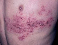
Bulla (pl. Bullae) - A raised, circumscribed lesion greater than 0.5 cm that contains serous fluid.
Nodule - A palpable, solid lesion, less than 1 cm in diameter. These are usually found in the dermal or subcutaneous tissue, and the lesion may be above, level with, or below the skin surface.
Tumor - Solid, firm lesions >1 cm that can be above, level with, or beneath the skin surface. Also known as a mass.
Wheal - Transient, circumscribed, elevated papules or plaques, often with erythematous borders and pale centers.
Pustule - A small (<>
Cataract: A waterfall that curtains your sight

What is a cataract?
When cataracts are mentioned, people often think of a film that grows on their eyes causing them to see double or blurred images. However, a cataract does not form on the eye, but rather within the eye.
Eye without a cataract
Eye with a cataract
A cataract is a clouding of the natural lens, the part of the eye responsible for focusing light and producing clear, sharp images. The lens is contained in a sealed bag or capsule. As old cells die they become trapped within the capsule. Over time, the cells accumulate causing the lens to cloud, making images look blurred or fuzzy. For most people, cataracts are a natural result of aging.
In fact, they are the leading cause of visual loss among adults 55 and older. Eye injuries, certain medications, and diseases such as diabetes and alcoholism have also been known to cause cataracts.

Watch Video: 8 minutes
Click on the Image
Subscribe to:
Posts (Atom)