Showing posts with label Infectious disease. Show all posts

Malaria : Epidemiology, signs and symptoms

FActs about Malaria
  1. Malaria is a life-threatening disease caused by parasites that are transmitted to people through the bites of infected mosquitoes.
  2. A child dies of malaria every 30 seconds.
  3. There were 247 million cases of malaria in 2006, causing nearly one million deaths, mostly among African children.
  4. Malaria is preventable and curable.
  5. Approximately half of the world's population is at risk of malaria, particularly those living in lower-income countries.
  6. Travellers from malaria-free areas to disease "hot spots" are especially vulnerable to the disease.Malaria takes an economic toll - cutting economic growth rates by as much as 1.3% in countries with high disease rates.

Malaria is caused by parasites of the species Plasmodium. The parasites are spread to people through the bites of infected mosquitoes.


There are four types of human malaria:

Plasmodium falciparum
Plasmodium vivax
Plasmodium malariae
Plasmodium ovale.
Plasmodium falciparum and Plasmodium vivax are the most common. Plasmodium falciparum is the most deadly.


Common symptoms of malaria
In the early stages, malaria symptoms are sometimes similar to those of many other infections caused by bacteria, viruses, or parasites. Symptoms may include:
Fever.
Chills.
Headache.
Sweats.
Fatigue.
Nausea and vomiting.
Other common symptoms of malaria include:
Dry (nonproductive) cough.
Muscle and/or back pain.
Enlarged Spleen

Complications of Malaria( Falciparum):
Cerebral malaria
Algid Malaria
ARDS
Convulsion
Coma
Acute Renal Failure
Hypoglycemia
hyperthermia
hyperparasitemia
severe anemia


Symptomatic diagnosis
Using Giemsa-stained blood smears from children in Malawi, one study showed that when clinical predictors (rectal temperature, nailbed pallor, and splenomegaly) were used as treatment indications, rather than using only a history of subjective fevers, a correct diagnosis increased from 21% to 41% of cases and unnecessary treatment for malaria was significantly decreased.

Microscopic examination of blood films
Field tests
In areas where microscopy is not available, or where laboratory staff are not experienced at malaria diagnosis, there are antigen detection tests that require only a drop of blood. Immunochromatographic tests (also called: Malaria Rapid Diagnostic Tests, Antigen-Capture Assay or "Dipsticks") have been developed, distributed and fieldtested. These tests use finger-stick or venous blood, the completed test takes a total of 15–20 minutes, and a laboratory is not needed. The threshold of detection by these rapid diagnostic tests is in the range of 100 parasites/µl of blood compared to 5 by thick film microscopy. The first rapid diagnostic tests were using P. falciparum glutamate dehydrogenase as antigen.[42] PGluDH was soon replaced by P.falciparum lactate dehydrogenase, a 33 kDa oxidoreductase [EC 1.1.1.27]. It is the last enzyme of the glycolytic pathway, essential for ATP generation and one of the most abundant enzymes expressed by P.falciparum. PLDH does not persist in the blood but clears about the same time as the parasites following successful treatment. The lack of antigen persistence after treatment makes the pLDH test useful in predicting treatment failure. In this respect, pLDH is similar to pGluDH. The OptiMAL-IT assay can distinguish between P. falciparum and P. vivax because of antigenic differences between their pLDH isoenzymes. OptiMAL-IT will reliably detect falciparum down to 0.01% parasitemia and non-falciparum down to 0.1%. Paracheck-Pf will detect parasitemias down to 0.002% but will not distinguish between falciparum and non-falciparum malaria. Parasite nucleic acids are detected using polymerase chain reaction. This technique is more accurate than microscopy. However, it is expensive, and requires a specialized laboratory. Moreover, levels of parasitemia are not necessarily correlative with the progression of disease, particularly when the parasite is able to adhere to blood vessel walls. Therefore more sensitive, low-tech diagnosis tools need to be developed in order to detect low levels of parasitaemia in the field. Areas that cannot afford even simple laboratory diagnostic tests often use only a history of subjective fever as the indication to treat for malaria. Using Giemsa-stained blood smears from children in Malawi, one study showed that unnecessary treatment for malaria was significantly decreased when clinical predictors (rectal temperature, nailbed pallor, and splenomegaly) were used as treatment indications, rather than the current national policy of using only a history of subjective fevers (sensitivity increased from 21% to 41%).


Molecular methods
Molecular methods are available in some clinical laboratories and rapid real-time assays (for example, QT-NASBA based on the polymerase chain reaction) are being developed with the hope of being able to deploy them in endemic areas.

Rapid antigen tests
OptiMAL-IT will reliably detect falciparum down to 0.01% parasitemia and non-falciparum down to 0.1%. Paracheck-Pf will detect parasitemias down to 0.002% but will not distinguish between falciparum and non-falciparum malaria. Parasite nucleic acids are detected using polymerase chain reaction. This technique is more accurate than microscopy. However, it is expensive, and requires a specialized laboratory. Moreover, levels of parasitemia are not necessarily correlative with the progression of disease, particularly when the parasite is able to adhere to blood vessel walls. Therefore more sensitive, low-tech diagnosis tools need to be developed in order to detect low levels of parasitaemia in the field.

Classification: Pneumonia

Early classification schemes


Initial descriptions of pneumonia focused on the anatomic or pathologic appearance of the lung, either by direct inspection at autopsy or by its appearance under a microscope.

A lobar pneumonia is an infection that only involves a single lobe, or section, of a lung. Lobar pneumonia is often due to Streptococcus pneumoniae (though Klebsiella pneumoniae is also possible.)
Multilobar pneumonia involves more than one lobe, and it often causes a more severe illness.
Bronchial pneumonia affects the lungs in patches around the tubes (bronchi or bronchioles).
Interstitial pneumonia involves the areas in between the alveoli, and it may be called "interstitial pneumonitis." It is more likely to be caused by viruses or by atypical bacteria.

The discovery of x-rays made it possible to determine the anatomic type of pneumonia without direct examination of the lungs at autopsy and led to the development of a radiological classification. Early investigators distinguished between typical lobar pneumonia and atypical (e.g. Chlamydophila) or viral pneumonia using the location, distribution, and appearance of the opacities they saw on chest x-rays. Certain x-ray findings can be used to help predict the course of illness, although it is not possible to clearly determine the microbiologic cause of a pneumonia with x-rays alone.

With the advent of modern microbiology, classification based upon the causative microorganism became possible. Determining which microorganism is causing an individual's pneumonia is an important step in deciding treatment type and length. Sputum cultures, blood cultures, tests on respiratory secretions, and specific blood tests are used to determine the microbiologic classification. Because such laboratory testing typically takes several days, microbiologic classification is usually not possible at the time of initial diagnosis.

Combined clinical classification
Traditionally, clinicians have classified pneumonia by clinical characteristics, dividing them into "acute" (less than three weeks duration) and "chronic" pneumonias. This is useful because chronic pneumonias tend to be either non-infectious, or mycobacterial, fungal, or mixed bacterial infections caused by airway obstruction. Acute pneumonias are further divided into the classic bacterial bronchopneumonias (such asStreptococcus pneumoniae), the atypical pneumonias (such as the interstitial pneumonitis of Mycoplasma pneumoniae or Chlamydia pneumoniae), and the aspiration pneumonia syndromes.

Chronic pneumonias, on the other hand, mainly include those of Nocardia, Actinomyces and Blastomyces dermatitidis, as well as the granulomatous pneumonias (Mycobacterium tuberculosis and atypical mycobacteria, Histoplasma capsulatum and Coccidioides immitis).

The combined clinical classification, now the most commonly used classification scheme, attempts to identify a person's risk factors when he or she first comes to medical attention. The advantage of this classification scheme over previous systems is that it can help guide the selection of appropriate initial treatments even before the microbiologic cause of the pneumonia is known. There are two broad categories of pneumonia in this scheme: community-acquired pneumonia and hospital-acquired pneumonia. A recently introduced type of healthcare-associated pneumonia (in patients living outside the hospital who have recently been in close contact with the health care system) lies between these two categories.

Community-acquired pneumonia
Community-acquired pneumonia (CAP) is infectious pneumonia in a person who has not recently been hospitalized. CAP is the most common type of pneumonia. The most common causes of CAP vary depending on a person's age, but they include Streptococcus pneumoniae, viruses, the atypical bacteria, and Haemophilus influenzae. Overall, Streptococcus pneumoniae is the most common cause of community-acquired pneumonia worldwide. Gram-negative bacteria cause CAP in certain at-risk populations. CAP is the fourth most common cause of death in the United Kingdom and the sixth in the United States. The term "walking pneumonia" has been used to describe a type of community-acquired pneumonia of less severity (because of the fact that the sufferer can continue to "walk" rather than require hospitalization). Walking pneumonia is usually caused by the atypical bacteria mycoplasma pneumonia.

Hospital-acquired pneumonia
Hospital-acquired pneumonia, also called nosocomial pneumonia, is pneumonia acquired during or after hospitalization for another illness or procedure with onset at least 72 hrs after admission. The causes, microbiology, treatment and prognosis are different from those of community-acquired pneumonia. Up to 5% of patients admitted to a hospital for other causes subsequently develop pneumonia. Hospitalized patients may have many risk factors for pneumonia, including mechanical ventilation, prolonged malnutrition, underlying heart and lung diseases, decreased amounts of stomach acid, and immune disturbances. Additionally, the microorganisms a person is exposed to in a hospital are often different from those at home . Hospital-acquired microorganisms may include resistant bacteria such as MRSA, Pseudomonas, Enterobacter, andSerratia. Because individuals with hospital-acquired pneumonia usually have underlying illnesses and are exposed to more dangerous bacteria, it tends to be more deadly than community-acquired pneumonia.Ventilator-associated pneumonia (VAP) is a subset of hospital-acquired pneumonia. VAP is pneumonia which occurs after at least 48 hours of intubation and mechanical ventilation.

Other types of pneumonia
Severe acute respiratory syndrome (SARS)
SARS is a highly contagious and deadly type of pneumonia which first occurred in 2002 after initial outbreaks in China. SARS is caused by the SARS coronavirus, a previously unknown pathogen.
Bronchiolitis obliterans organizing pneumonia (BOOP)
BOOP is caused by inflammation of the small airways of the lungs. It is also known as cryptogenic organizing pneumonitis (COP).
Eosinophilic pneumonia
Eosinophilic pneumonia is invasion of the lung by eosinophils, a particular kind of white blood cell. Eosinophilic pneumonia often occurs in response to infection with a parasite or after exposure to certain types of environmental factors.
Chemical pneumonia
Chemical pneumonia (usually called chemical pneumonitis) is caused by chemical toxicants such as pesticides, which may enter the body by inhalation or by skin contact. When the toxic substance is an oil, the pneumonia may be called lipoid pneumonia.
Aspiration pneumonia
Aspiration pneumonia (or aspiration pneumonitis) is caused by aspirating foreign objects which are usually oral or gastric contents, either while eating, or after reflux or vomiting which results inbronchopneumonia. The resulting lung inflammation is not an infection but can contribute to one, since the material aspirated may contain anaerobic bacteria or other unusual causes of pneumonia. Aspiration is a leading cause of death among hospital and nursing home patients, since they often cannot adequately protect their airways and may have otherwise impaired defenses.

Dust pneumonia
Dust pneumonia describes disorders caused by excessive exposure to dust storms, particularly during the Dust Bowl in the United States. With dust pneumonia, dust settles all the way into the alveoli of the lungs, stopping the cilia from moving and preventing the lungs from ever clearing themselves.

Necrotizing pneumonia, although overlapping with many other classifications, includes pneumonias that cause substantial necrosis of lung cells, and sometimes even lung abscess. Implicated bacteria are extremely commonly anaerobic bacteria, with or without additional facultatively anaerobic ones like Staphylococcus aureus, Klebsiella pneumoniae and Streptococcus pyogenes. Type 3 pneumococcus is uncommonly implicated.

Opportunistic pneumonia includes those that frequently strike immunocompromised victims. Main pathogens are cytomegalovirus, Pneumocystis jiroveci, Mycobacterium avium-intracellulare, invasiveaspergillosis, invasive candidiasis, as well as the "usual bacteria" that strike immunocompetent people as well.

Swine Flu 4 vaccines Declared

The US Food and Drug Administration (FDA) announced today that it has approved 4 vaccines against the 2009 influenza A (H1N1) virus, formerly known as "swine flu." The vaccine lots are expected to be available and distributed within the next 4 weeks.

FDA Commissioner of Food and Drugs Margaret A. Hamburg, MD, said she thought Tuesday's approval was good news for the nation's response to the H1N1 influenza virus. "This vaccine will help protect individuals from serious illness and death from influenza," she said.

The approval comes at a time when the Centers for Disease Control and Prevention (CDC) is reporting that visits to physicians around the country for influenza-like illness are increasing and are higher than expected at this time of year. The vaccines that are currently available against 3 seasonal influenza virus strains will not protect against the 2009 H1N1 virus.

The FDA said that the vaccines, based on early data, effectively elicit an immune response in most healthy adults about 8 to10 days after vaccination. Clinical studies are still underway to produce an optimal dose for children, with results expected in the near future.

Meanwhile, the CDC stresses that influenza is primarily spread through person-to-person contact, by the coughing or sneezing of infected people, and recommends that infected people stay home and limit their contact with others to keep from infecting them.

The newly approved vaccines are being made by CSL Limited, MedImmune LLC, Novartis Vaccines and Diagnostics Limited, and Sanofi Pasteur Inc. All 4 firms reportedly use the same processes to manufacture the H1N1 vaccines. As with the seasonal influenza vaccine, some lots of the H1N1 vaccine will contain the preservative thimerosal and others will not. The FDA has been continuing its efforts toward reducing thimerosal used in vaccines.

The FDA warns that persons with known allergies to chicken eggs or any other substance in the vaccine should probably not be vaccinated, although in the ongoing clinical trials, the vaccines have been well tolerated. The most common adverse effect is soreness at the injection site; other adverse effects can include a mild fever, body aches, and fatigue for a couple of days after vaccination. For the nasal spray delivery system, the most common adverse effects were runny nose, nasal congestion in all ages, sore throats in adults, and fever in children aged 2 to 6 years.

The FDA is working with different organizations regarding adverse event monitoring, information sharing, and an overall analysis during and after the 2009 H1N1 vaccination program, according to the news release. "As with any medical product, unexpected or rare serious adverse events may occur," the FDA notes.

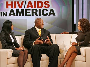
Magic Johnson: Fight Against HIV/AIDS


Magic Johnson: Personal life and His Commitment against AIDS

Personal life

Johnson first fathered a son in 1981, when Andre Johnson was born to Melissa Mitchell. Although Andre was raised by his mother, he visited Johnson each summer, and as of October 2005 was working for Magic Johnson Enterprises as a marketing director. In 1991, Johnson married Earlitha "Cookie" Kelly, with whom he had one son, Earvin III; the couple adopted a daughter, Elisa, in 1995.

In 1998, Johnson hosted a late night talk show on the Fox network called The Magic Hour, but the show was canceled after two months due to low ratings. He runs Magic Johnson Enterprises, a company that has a net worth of $700 million; its subsidiaries include Magic Johnson Productions, a promotional company; Magic Johnson Theaters, a nationwide chain of movie theaters; and Magic Johnson Entertainment, a movie studio. Johnson has also worked as a motivational speaker. He is a supporter of the Democratic Party—in 2005, he publicly endorsed Phil Angelides for governor of California, and Hillary Clinton for president of the United States. Johnson was an NBA commentator for Turner Network Television for seven years, before becoming a studio analyst for ESPN's NBA Countdown in 2008.

After announcing his infection in November 1991, Johnson created the Magic Johnson Foundation to help combat HIV, although he later diversified the foundation to include other charitable goals. In 1992, he joined the National Commission on AIDS, but left after eight months, saying that the commission was not doing enough to combat the disease. He was also the main speaker for the United Nations (UN) World AIDS Day Conference in 1999, and has served as a United Nations Messenger of Peace.

HIV had been associated with drug addicts and homosexuals, but Johnson's campaigns sought to show that the risk of infection was not limited to those groups. Johnson stated that his aim was to "help educate all people about what [HIV] is about" and teach others not to "discriminate against people who have HIV and AIDS". Johnson was later criticized by the AIDS community for his decreased involvement in publicizing the spread of the disease.

To prevent his HIV infection from becoming AIDS, Johnson takes a daily combination of drugs from GlaxoSmithKline and Abbott Laboratories. He has advertised GlaxoSmithKline's drugs,[91] and partnered with Abbott Laboratories to publicize the fight against AIDS in African American communities.

WHO Issues Guidelines for Antiviral Treatment for H1N1 and Other Influenza

WHO Issues Guidelines for Antiviral Treatment for H1N1 and Other Influenza

August 25, 2009 —
The World Health Organization (WHO) has issued guidelines for antiviral treatment for novel influenza A (H1N1) and other influenza. The purpose of the new recommendations, which were posted online August 20, is to provide a basis for advice to clinicians regarding the use of the currently available antivirals for patients presenting with illness caused by influenza virus infection, as well as considerations regarding potential use of these antiviral medications for chemoprophylaxis.

On the basis of a review of data collected with previously circulating strains, and treatment of human H5N1 influenza virus infections, the new guidelines expand on recommendations published in May 2009, titled ʺClinical management of human infection with new influenza A (H1N1) virus: Initial guidance." These new guidelines do not change recommendations in the WHO rapid advice guidelines on pharmacological management of humans infected with highly pathogenic avian influenza A (H5N1) virus.

"In April 2009, the [WHO] received reports of sustained person to person infections with [H1N1] virus in Mexico and the United States," write Edgar Bautista, from Médico Neumólogo Intensivista, Jefe de UCI-INER in Mexico, and colleagues. "Subsequent international spread led WHO to declare on 11 June 2009 that the first influenza pandemic in 41 years had occurred. This 2009 pandemic H1N1 influenza virus has now spread worldwide, with confirmed cases of pandemic H1N1 virus infection reported in more than 100 countries in all 6 WHO regions[, which] has led to the need to add to the existing guidance on the use of antivirals."

The new recommendations highlight oseltamivir and zanamivir, which are neuraminidase inhibitors, and amantadine and rimantadine, which are M2 inhibitors. Suggestions are also provided regarding the use of some other potential pharmacological treatments, such as ribavirin, interferons, immunoglobulins, and corticosteroids.

Management of patients with pandemic influenza (H1N1) 2009 virus infection is the primary focus of the statement, although it also includes guidance regarding the use of the antivirals for treatment of other seasonal influenza virus strains, as well as for infections resulting from novel influenza A virus strains.

The guidelines urge country and local public health authorities to issue local recommendations for clinicians periodically, based on epidemiological and antiviral susceptibility data on the locally circulating influenza strains. As the prevalence and severity of the current pandemic evolves, WHO anticipates that additional data will be forthcoming that may require revision of the current recommendations. WHO therefore plans to review the guidance no later than September 2009 to determine whether modifications to the recommendations are needed.

Recommendations for Antiviral Treatment of H1N1

For patients with confirmed or strongly suspected infection with influenza pandemic (H1N1) 2009, when antiviral medications for influenza are available, specific recommendations regarding use of antivirals for treatment of pandemic (H1N1) 2009 influenza virus infection are as follows:

•Oseltamivir should be prescribed, and treatment started as soon as possible, for patients with severe or progressive clinical illness (strong recommendation, low-quality evidence). Depending on clinical response, higher doses of up to 150 mg twice daily and longer duration of treatment may be indicated. This recommendation is intended for all patient groups, including pregnant women, neonates, and children younger than 5 years of age.
•Zanamivir is indicated for patients with severe or progressive clinical illness when oseltamivir is not available or not possible to use, or when the virus is resistant to oseltamivir but known or likely to be susceptible to zanamivir (strong recommendation, very low quality evidence).
•Antiviral treatment is not required in patients not in at-risk groups who have uncomplicated illness caused by confirmed or strongly suspected influenza virus infection (weak recommendation, low-quality evidence). Patients considered to be at risk are infants and children younger than 5 years of age; adults older than 65 years of age; nursing home residents; pregnant women; patients with chronic comorbid disease including cardiovascular, respiratory, or liver disease and diabetes; and immunosuppressed patients because of malignancy, HIV infection, or other diseases.
•Oseltamivir or zanamivir treatment should be started as soon as possible after the onset of illness in patients in at-risk groups who have uncomplicated illness caused by influenza virus infection (strong recommendation, very low quality evidence).
Recommendations for Chemoprophylaxis of H1N1

Specific recommendations regarding the use of antivirals for chemoprophylaxis of pandemic (H1N1) 2009 influenza virus infection are as follows:

•When risk for human-to-human transmission of influenza is high or low, and the probability of complications of infection is high, either because of the influenza strain or because of the baseline risk of the exposed group, use of oseltamivir or zanamivir may be considered as postexposure chemoprophylaxis for the affected community or group, for individuals in at-risk groups, or for healthcare workers (weak recommendation, moderate-quality evidence).
•Individuals in at-risk groups or healthcare personnel need not be offered antiviral chemoprophylaxis if the likelihood of complications of infection is low. This recommendation should be applied independent of risk for human-to-human transmission (weak recommendation, low-quality evidence).
For treatment of mild to moderate uncomplicated clinical presentation of infection with multiple cocirculating influenza A subtypes or viruses with different antiviral susceptibilities, patients in at-risk groups should be treated with zanamivir or oseltamivir plus M2 inhibitor (noting that amantadine should not be used in pregnant women). Otherwise-healthy patients with this presentation need not be treated.

When the clinical presentation of infection with multiple cocirculating influenza A subtypes or viruses with different antiviral susceptibilities is severe or progressive, all patients should be treated with oseltamivir plus M2 inhibitor, or zanamivir.

For treatment of mild to moderate uncomplicated clinical presentation of infection with sporadic zoonotic influenza A viruses including H5N1, the at-risk population should be treated with oseltamivir or zanamivir, and the otherwise-healthy population with oseltamivir. All patients, regardless of risk status, with severe or progressive presentation of infection with sporadic zoonotic influenza A viruses including H5N1 should be treated with oseltamivir plus an M2 inhibitor.

((News From MEDSCAPE NEWS))

Specific Virus Families causing disease in animals and plants


  1. Adenovirus

  2. Afican Swine Fever Virus

  3. Animal Viruses

  4. Arbovirus

  5. Arenavirus
  6. Arterivirus
  7. Astrovirus
  8. Bacteriophage
  9. Baculovirus

  10. Bunyavirus

  11. Calicivirus

  12. Caulimovirus

  13. Coronavirus
  14. Filovirus

  15. Flavivirus

  16. Hepadnavirus

  17. Herpesvirus
  18. Myovirus

  19. Nodavirus

  20. Orthomyxovirus

  21. Paramyxovirus

  22. Papovavirus

  23. Parvovirus

  24. Phycodnavirus

  25. Picornavirus

  26. Poxvirus
  27. Reovirus

  28. Retrovirus

  29. Rhabdovirus

  30. Togavirus
  31. Viroids

Swine Flu : Who Are at Risk

Which groups are at greatest risk?

Some people are more at risk than others of serious illness if they catch swine flu. They will need to start taking antivirals as soon as they are confirmed with the illness. On occasion, doctors may advise some high risk patients to take antivirals before they have symptoms if someone close to them has swine flu.

The risk profile of the virus is still being studied but it is already known that certain groups of people are particularly vulnerable. These include:

  • Patients who have had drug treatment for asthma in the past three years
  • Pregnant women
  • People aged 65 years and older
  • Children under five years old
  • People with chronic lung disease
  • People with chronic heart disease
  • People with chronic kidney disease
  • People with chronic liver disease
  • People with chronic neurological disease
  • People with immunosuppression (whether caused by disease or treatment)
  • People with diabetes mellitus

Why are healthy people over 65 and children not a priority for the swine flu vaccine?

Healthy people aged over 65 appear to have some natural immunity to the swine flu virus. And while children are disproportionately affected by swine flu, the vast majority make a full recovery - therefore the experts do not advise that children (other than those in at-risk groups) should be vaccinated initially.

When Did AIDS Begin? How?


When Did AIDS

Begin?

Spring 1988
A new study of the oldest known HIV suggests the virus jumped from animals to humans in the 1940s.

The year was 1959, location: The central African city of Leopoldville, now called Kinshasa, shortly before the waves of violent rebellion that followed the liberation of the Belgian Congo. A seemingly healthy man walked into a hospital clinic to give blood for a Western backed study of blood diseases. He walked away and was never heard from again. Doctors analyzed his sample, froze it in a test tube and forgot about it. A quarter-century later, in the mid-1980s, researchers studying the growing AIDS epidemic took a second look at the blood and discovered that it contained HIV, the virus that causes AIDS.

And not just any HIV. The Leopoldville sample is the oldest specimen of the AIDS virus ever isolated and may now help solve the mystery of how and when the virus made the leap from animals (monkeys or chimpanzees) to humans, according to a report published last week in Nature. Dr. David Ho, director of the Aaron Diamond AIDS Research Center in New York City and one of the study's authors, says a careful genetic analysis of the sample's DNA pushes the origin of the AIDS epidemic back at least a decade, to the early '50s or even the '40s.

Over the past 15 years, scientists have identified at least 10 subtypes of the virus. But they couldn't tell whether they were seeing variations on one changeable virus or the handiwork of several different viruses that had made the jump from primates to man. A look at the genetic mutations in the Leopoldville sample strongly suggests that all it took to launch the epidemic was one unlucky turn of events.

By comparing the DNA of the 1959 virus with that of samples taken from the '80s and '90s. Ho and his colleagues constructed a viral family tree in which the Leopoldville isolate sits right at the juncture where three subtypes branch out. The 39-year-old specimen is also strikingly similar to the other seven subtypes. The clear implication: all the viral strains can be traced back to a single event or a closely related group of events. One theory is that AIDS started through contact with infected monkeys in a remote area and spread to the rest of the population through urbanization and mass inoculations.

The findings underscore how rapidly HIV can adapt to its surroundings, making it difficult to develop effective vaccines. No one knows how many more subtypes of HIV will sprout in the next 40 years, but chances are they will be every bit as lethal as the ones we see today, if not more so.

Swine Flu :How to Protect yourself


preventive measure
s for Swine flu—

1. The first preventive measure is to avoid contact with the pigs (swine). If you have pigs in your area then please inform the local municipal office so that he can take care of those pigs by keeping them isolated.

2. Swine flu is communicable disease, so
use the face masks to protect from the swine flu antigens.

3.
Cover your nose and mouth when coughing or sneezing, using tissue when possible. Dispose this tissue by using only once.

4.
Avoid visiting the crowded places like theaters and prayer halls. This can be the spreading ground for Swine flu

5.
Maintain good hygiene. Wash your hands frequently with soap and water to reduce the spread of virus. It would be better if you use alcohol sanitizers or Dettol for washing hands.

6. Take a special care of children because they easily get infected with the Swine flu. It is okay if you don’t send them to school for few days. Many schools have even announced holidays.

7.
Avoid eating outside food because it may be contaminated and may make you infected with the virus.

8.
Don’t use the public urinals because many people spit there, which could lead to the spreading of the disease.

9. Drink the boiled water.

Anthrax: Disease or A Biological Weapon

How the Anthrax Spreads.


ANTHRAX: Danger we face


Anthrax is a deadly disease caused by the bacteria Bacillus anthracis. This disease has animal origin and mainly the victims are the grazing animals. However, the disease can spread to humans, who come in contact either with the affected animals or their burial grounds. Anthrax is not a contagious disease and does not spread from person toperson. This disease is known to spread from animals or by bioterrorism
.

Anthrax bacteria have long life and can survive in soil for many years. Wild or domestic animals that graze in or around the soil can get infected while eating rough or irritant vegetation. Such vegetation when eaten causes wound within the gastrointestinal tract and this causes the bacteria to enter into the tissues. The bacteria start multiplying and begin producing toxins which finally leads to death.

Exposure to infected animals or their products such as skin, wool or meat is the route taken by the bacteria to enter the human body. Workers, who work with dead animals or their products, are at a greater risk to this disease. People who are exposed to low levels of these bacteria may not develop the infection as the body’s defense mechanism is strong enough to withstand it. A dangerous form of anthrax through inhalation while sorting wool was prevalent long back and this was known as Wool Sorter’s disease. Now such infections are very rare as the infected animals are not available any more.

Anthrax pores have been used as biological weapons of mass destruction. The last reported use was by the Rhodesian government against cattle and humans during its war with black nationalists in 1978-1979. Anthrax was deliberately spread using the postal system in United States in 2001. This caused 21 cases of Anthrax infection.

Anthrax infection can come through three routes. Through the skin, while handling products of infected animals; through inhalation by inhaling anthrax pores from animal products; or through gastrointestinal path by eating undercooked meat from infected animals.

Swine Flu Alert : India And Nepal

Swine Flu Now In South Asia

http://www.topnews.in/files/india-swine.jpg

Current Pandemic Level : 6


The WHO has raised the Influenza Pandemic Alert to the highest level which is 6. Already India has seen over 40 confirmed cases of Swine Flu and the threat of a full blown epidemic in India is very real. The best we citizens can do is keep ourselves informed about the happenings and the steps we can take to prevent the spread of the flu. With most of the affected people fitting the profile of a person with internet access, we believe the internet is the first place that people would come looking for information like symptoms, who to contact etc. Swine Flu India is an attempt to bring all the necessary information in one place. India cannot afford to fall sick. Keep yourself and others informed.


Some Vital Stats


Total Confirmed Cases in India


959**

Total Deaths in India


10**

Total Confirmed Cases Worldwide


162380*

Total Deaths Worldwide


1154*

Last Updated Date and Time


11/08/2009 19:26 IST

Sources : *WHO **MoHFW


Nepal = 20 cases Reported

Swine Flu : CDC Recommendation


Novel H1N1 Vaccination Recommendations

With the new H1N1 virus continuing to cause illness, hospitalizations and deaths in the US during the normally flu-free summer months and some uncertainty about what the upcoming flu season might bring, CDC's Advisory Committee on Immunization Practices has taken an important step in preparations for a voluntary novel H1N1 vaccination effort to counter a possibly severe upcoming flu season. On July 29, ACIP met to consider who should receive novel H1N1 vaccine when it becomes available.

Novel H1N1 Vaccine

Every flu season has the potential to cause a lot of illness, doctor’s visits, hospitalizations and deaths. CDC is concerned that the new H1N1 flu virus could result in a particularly severe flu season this year. Vaccines are the best tool we have to prevent influenza. CDC hopes that people will start to go out and get vaccinated against seasonal influenza as soon as vaccines become available at their doctor’s offices and in their communities (this may be as early as August for some). The seasonal flu vaccine is unlikely to provide protection against novel H1N1 influenza. However a novel H1N1 vaccine is currently in production and may be ready for the public in the fall. The novel H1N1 vaccine is not intended to replace the seasonal flu vaccine – it is intended to be used along-side seasonal flu vaccine.

CDC’s Advisory Committee on Immunization Practices (ACIP), a

panel made up of medical and public health experts, met July 29, 2009, to make recommendations on who should receive the new H1N1 vaccine when it becomes available. While some issues are still unknown, such as how severe the virus will be during the fall and winter months, the ACIP considered several factors, including current disease patterns, populations most at-risk for severe illness based on current trends in illness, hospitalizations and deaths, how much vaccine is expected to be available, and the timing of vaccine availability.

The groups recommended to receive the novel H1N1 influenza vaccine include:

  • Pregnant women because they are at higher risk of complications and can potentially provide protection to infants who cannot be vaccinated;
  • Household contacts and caregivers for children younger than 6 months of age because younger infants are at higher risk of influenza-related complications and cannot be vaccinated. Vaccination of those in close contact with infants less than 6 months old might help protect infants by “cocooning” them from the virus;
  • Healthcare and emergency medical services personnel because infections among healthcare workers have been reported and this can be a potential source of infection for vulnerable patients. Also, increased absenteeism in this population could reduce healthcare system capacity;
  • All people from 6 months through 24 years of age
    • Children from 6 months through 18 years of age because we have seen many cases of novel H1N1 influenza in children and they are in close contact with each other in school and day care settings, which increases the likelihood of disease spread, and
    • Young adults 19 through 24 years of age because we have seen many cases of novel H1N1 influenza in these healthy young adults and they often live, work, and study in close proximity, and they are a frequently mobile population; and,
  • Persons aged 25 through 64 years who have health conditions associated with higher risk of medical complications from influenza.

We do not expect that there will be a shortage of novel H1N1 vaccine, but flu vaccine availability and demand can be unpredictable and there is some possibility that initially, the vaccine will be available in limited quantities. So, the ACIP also made recommendations regarding which people within the groups listed above should be prioritized if the vaccine is initially available in extremely limited quantities. For more information see the CDC press release CDC Advisors Make Recommendations for Use of Vaccine Against Novel H1N1.

Once the demand for vaccine for the prioritized groups has been met at the local level, programs and providers should also begin vaccinating everyone from the ages of 25 through 64 years. Current studies indicate that the risk for infection among persons age 65 or older is less than the risk for younger age groups. However, once vaccine demand among younger age groups has been met, programs and providers should offer vaccination to people 65 or older.

SEARCH THIS BLOG FOR