
Apert Syndrome

 Introduction
Apert syndrome is a rare genetic disorder that is characterized by specific craniofacial and limb abnormalities. It is caused by a genetic mutation in the FGFR2 gene on chromosome 10. The mutation can be inherited from a parent who has Apert syndrome or it can be a spontaneous (new) mutation. Studies show that Apert syndrome tends to occur more often in children with older fathers. Furthermore, all new mutations (those that have not been inherited by an affected parent) have been shown to occur exclusively in the FGFR2 gene received by the father. Apert syndrome occurs in 1 out of 100,000 to 160,000 live births and affects males and females equally. The first reported case of the syndrome was in 1848 by S.W. Wheaton, and in 1906, a French physician named E. Apert described nine cases and defined the syndrome.
Features and Characteristics
The following characteristics have been found in children with Apert syndrome:
- Prematurely fused cranial sutures
- Retruded (or sunken) mid-face
- Fused fingers
- Fused toes
- Brachycephaly (short wide head)
- Acrocephaly (high prominent forehead)
- Flattened back of skull
- Prominent eyes - may be spaced widely apart or slant downward
- Strabismus
- Prominent mandible
- Depressed nasal bridge and small anteverted nose
- Down-turned corners of the mouth
- Low set ears (as well as hearing loss)
- Cleft palate
- Severe acne in teens
- Hydrocephalus
- Dental abnormalities (malposition of the teeth, crowding of the teeth, delayed tooth eruption, high-arched narrow palate, thickened ridges that support the teeth)
- Internal organ abnormalities including heart defects and abnormalities of the trachea,
- uterus, and brain
Skin manifestations of Apert syndromeHyperhidrosis
Synonychia
Brittle nails
Severe acne in puberty
Interruption of the eyebrows
Hypopigmentation
Hyperkeratosis
Paronychial infections of plantar skin
Excessive skin wrinkling of forehead
Dimples at knuckles, shoulders and elbows
|
Prematurely Fused Cranial Sutures
Retruded Mid-face
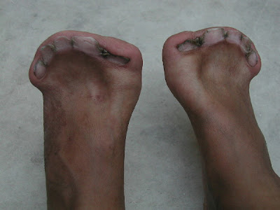
Fused Fingers and Toes
Diagnosis
The diagnosis can be made by a skull x-ray, which will confirm premature closure of the skull, and by a clinical exam. The combination of the craniofacial problems and the fused fingers and toes is what distinguishes Apert syndrome from other similar syndromes. Since the defect which causes Apert syndrome has been identified, genetic testing can be provided to confirm a diagnosis.
Treatment
Treating a child with Apert syndrome is best accomplished with a team approach. This would include a craniofacial surgeon, neurosurgeon, ENT specialist, audiologist, speech pathologist, oral surgeon, psychologist, ophthalmologist, and an orthodontist. The majority of treatment methods is surgical and the individual will likely require many operations. Aside from the surgeries required to correct the craniofacial problems and the fused fingers and toes, there may be other potential surgeries to improve the upper airway, address severe eye problems, or correct dental issues.
 Rickets : Valgus deformity ( knock Knee)
 Pot Belly in rickets
 Bowing of Legs: Genu Varum
  Widening of Wrist


